Copyright
©The Author(s) 2016.
World J Radiol. May 28, 2016; 8(5): 460-471
Published online May 28, 2016. doi: 10.4329/wjr.v8.i5.460
Published online May 28, 2016. doi: 10.4329/wjr.v8.i5.460
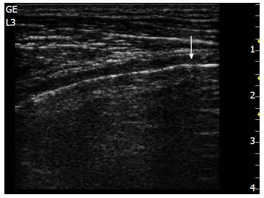
Figure 1 Longitudinal approach with longitudinal probe; allow to see upper and lower ribs visible (a), along with their shadow’s cone, and pleural line (arrow).
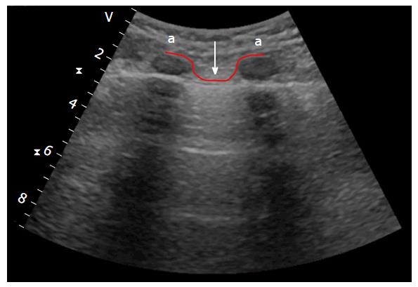
Figure 2 Bat sign with convex probe.
Consistsmake of two adjacent ribs with the pleural line between [upper and lower ribs are the “wings” of the “bat”(a), whileand, the pleural line that is the “back body” of the “bat” (arrow)]. Under Below the pleural line it is also possible to see two A-lines.
Figure 3 M-mode and seashore sign assist with, helps to the documentation of lung sliding on a picture.
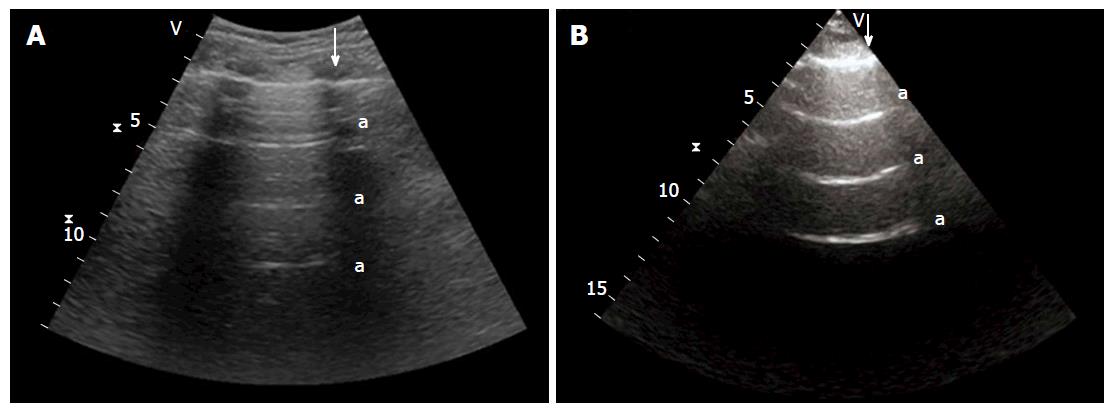
Figure 4 A-lines.
A: A-lines whit with convex probe; B: A-lines whit with sector probe. Arrow shows the pleural line, (a) shows A-lines.
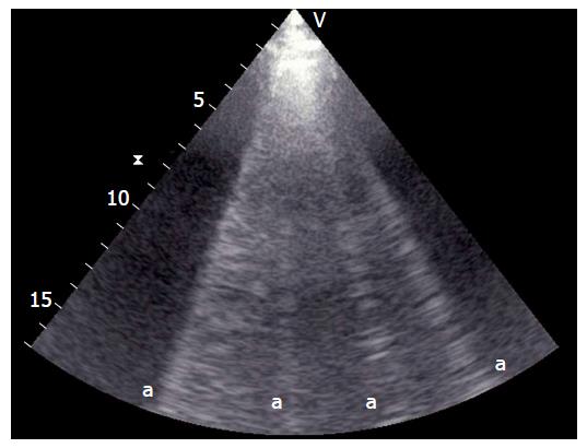
Figure 5 B-lines.
(a) shows B-lines.
Figure 6 Pleural effusion.
Pleural effusion is an echo-free [dark zone (a)], which and determinates the compression of the lung that appears consolidated, and floating in the pleural effusion (arrow).
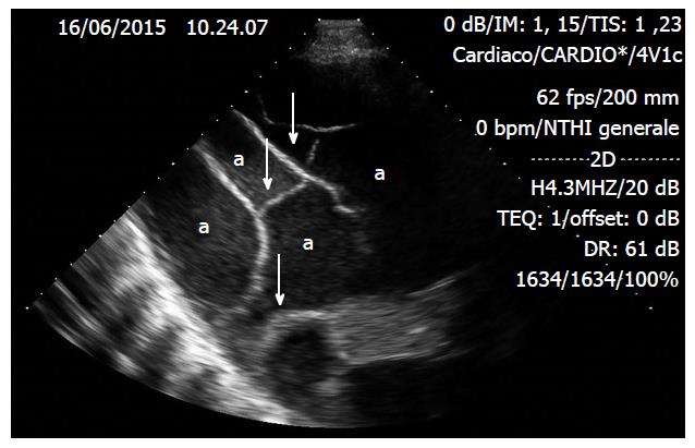
Figure 7 Loculated pleural effusion, which appears as a dark zone (a) and contains immobile septations (arrow) with encapsulated liquid.
Figure 8 Stratosphere sign.
Pointing M-mode into the zone characterized by the abolition lack of lung sliding it shows a characteristic pattern, namely the stratosphere sign (as opposed to the normal seashore sign) (by image courtesy of Dr. Ilario De Sio).
Figure 9 Lung point (arrow).
Is the precise area of the chest wall, where the regular reappearance of the lung sliding replaces the pneumothorax pattern and it corresponds to the point where the visceral and parietal pleura regain contact with each other (by image courtesy of Dr. Lucia Morelli).
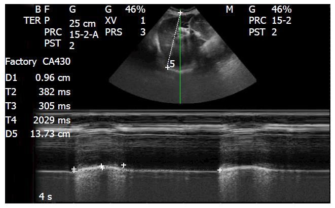
Figure 10 M-mode, diaphragmatic excursion (see the text) (by image courtesy of Dr.
Lucia Morelli).
- Citation: Liccardo B, Martone F, Trambaiolo P, Severino S, Cibinel GA, D’Andrea A. Incremental value of thoracic ultrasound in intensive care units: Indications, uses, and applications. World J Radiol 2016; 8(5): 460-471
- URL: https://www.wjgnet.com/1949-8470/full/v8/i5/460.htm
- DOI: https://dx.doi.org/10.4329/wjr.v8.i5.460


