Copyright
©The Author(s) 2016.
World J Radiol. Apr 28, 2016; 8(4): 403-409
Published online Apr 28, 2016. doi: 10.4329/wjr.v8.i4.403
Published online Apr 28, 2016. doi: 10.4329/wjr.v8.i4.403
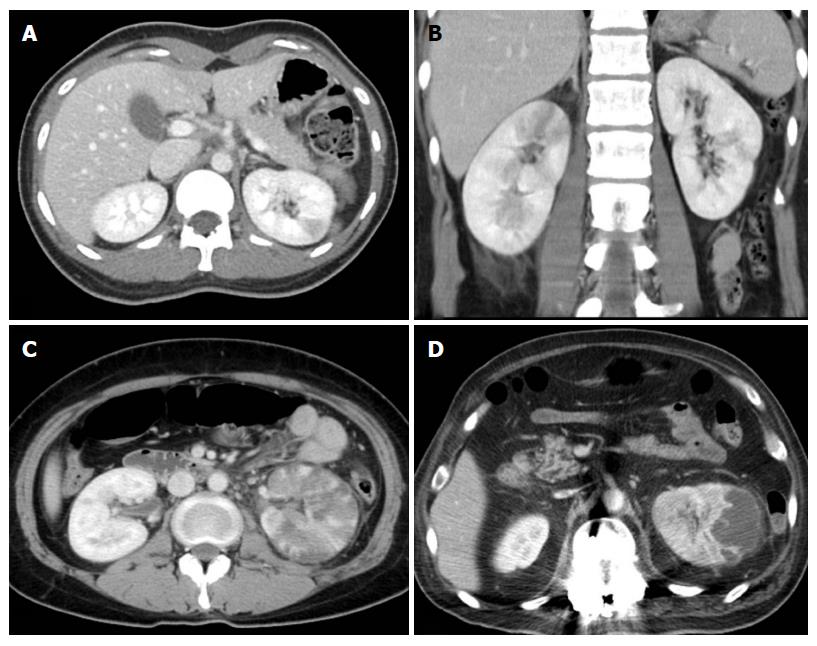
Figure 1 Grades of acute pyelonephritis.
A: Grade 1 APN in the left kidney that is defined as a focal, wedge-shape, hypodense lesion; B: Grade 2 APN in the right kidney that is defined as multifocal or diffuse hypodense lesions; C: Grade 3 acute pyelonephritis in the left kidney that is defined as abscesses less than 2 cm within the APN lesion; D: Grade 4 APN in the left kidney that is defined as large abscess pockets more than 2 cm in the longest diameter. APN: Acute pyelonephritis.
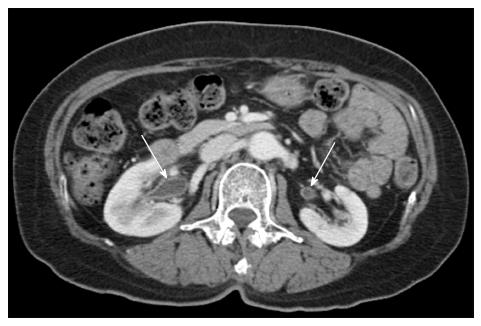
Figure 2 Urothelial thickening of bilateral renal pelvis (arrows) in a 64-year-old female with bacteremic acute pyelonephritis due to Escherichia coli.
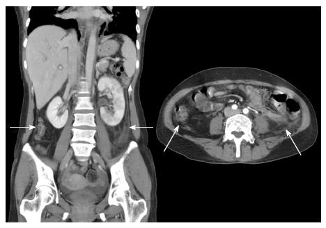
Figure 3 Diffuse peritoneal thickening (arrows) in a 42-year-old female with bacteremic acute pyelonephritis due to Escherichia coli.
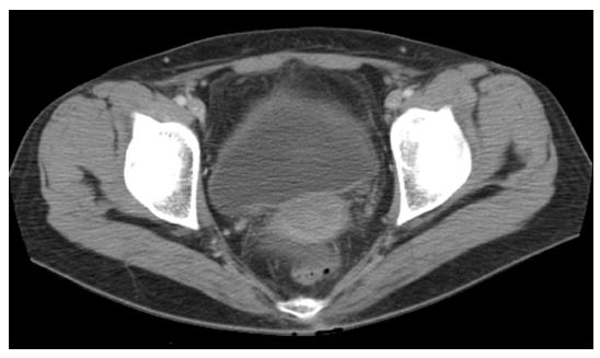
Figure 4 Infiltration into perivesical fat in a 31-year-old female with bacteremic acute pyelonephritis due to Escherichia coli.
- Citation: Oh SJ, Je BK, Lee SH, Choi WS, Hong D, Kim SB. Comparison of computed tomography findings between bacteremic and non-bacteremic acute pyelonephritis due to Escherichia coli. World J Radiol 2016; 8(4): 403-409
- URL: https://www.wjgnet.com/1949-8470/full/v8/i4/403.htm
- DOI: https://dx.doi.org/10.4329/wjr.v8.i4.403


