Copyright
©The Author(s) 2016.
World J Radiol. Dec 28, 2016; 8(12): 916-921
Published online Dec 28, 2016. doi: 10.4329/wjr.v8.i12.916
Published online Dec 28, 2016. doi: 10.4329/wjr.v8.i12.916
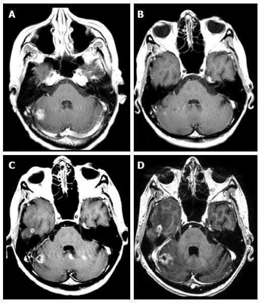
Figure 1 Follow-up axial enhanced T1-weighted magnetic resonance images of a brain metastasis from breast carcinoma treated with stereotactic radiosurgery in a 60-year-old woman.
A: Pre stereotactic radiosurgery (SRS) magnetic resonance (MR) image; B: Initial follow-up MR image at 6 wk after SRS demonstrating an initial volume reduction; C: Follow-up MR image at 9 wk after SRS demonstrating a transient volume increase (pseudo-progression); D: Follow-up MR image at 12 wk demonstrating a final volume reduction.
Figure 2 Follow-up axial enhanced T1-weighted magnetic resonance images of a lung carcinoma metastatic to the right cerebellum treated with stereotactic radiosurgery.
A: Pre stereotactic radiosurgery (SRS) magnetic resonance (MR) image; B: Initial follow-up MR image at 6 wk after SRS demonstrating an initial volume reduction; C: Follow-up MR image at 9 wk demonstrating volume increase (true-progression); D: Follow-up MR image at 12 wk with final volume increase.
- Citation: Sparacia G, Agnello F, Banco A, Bencivinni F, Anastasi A, Giordano G, Taibbi A, Galia M, Bartolotta TV. Value of serial magnetic resonance imaging in the assessment of brain metastases volume control during stereotactic radiosurgery. World J Radiol 2016; 8(12): 916-921
- URL: https://www.wjgnet.com/1949-8470/full/v8/i12/916.htm
- DOI: https://dx.doi.org/10.4329/wjr.v8.i12.916


