Copyright
©The Author(s) 2016.
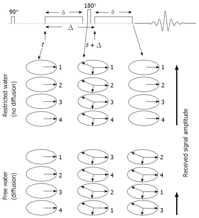
Figure 1 Schematic representation of the pulsed field gradient pulse sequence.
In this description we assume that we start the sequence with a sample containing only four in-phase spins labelled with 1, 2, 3 and 4. In the absence of diffusion, the first gradient pulse causes dephasing of the spins. The 180° radio-frequency pulse reverses the sign of the phase angle and thus after the second gradient pulse all spins are in phase which gives a maximum echo signal. In the presence of diffusion, spins go through a random walk process resulting in a distribution of phases. This in turn results in poorer refocusing of the spins and thus, a smaller echo signal.
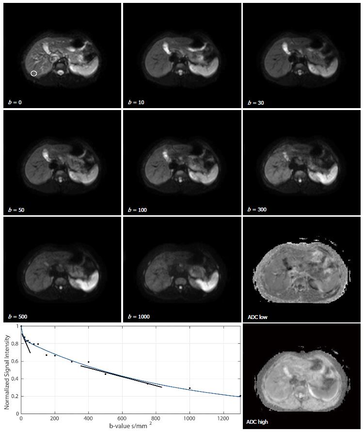
Figure 2 Diffusion-weighted magnetic resonance images of the abdomen of a healthy 25-year-old male volunteer at different b-values of 0, 10, 30, 50, 100, 300, 500, 1000 s/mm2.
An ROI placed over a non-heterogeneous region in the liver is shown on the b = 0 s/mm2 image. A bi-exponential fit to the ROI drawn on the diffusion-weighted-magnetic resonance data acquired with b-values of 0, 10, 20, 30, 40, 50, 75, 100, 150, 200, 300, 400, 500, 750, 1000 and 1300 s/mm2 is also shown where the slopes of the exponents represent the fast diffusion component (which includes perfusion) and the slower diffusion component. Quantitative apparent diffusion coefficients maps are also shown where ADC low was computed with b-values ≤ 100 s/mm2 and ADC high was computed with b-values ≥ 150 s/mm2. ROI: Region-of-interest; ADC: Apparent diffusion coefficient.
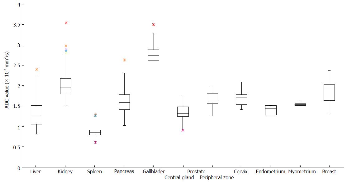
Figure 3 Box and whisker plots of the summarised apparent diffusion coefficient values reported for extra-cranial organs.
A total of 115 studies were summarised including for liver parenchyma, kidney (renal parenchyma), pancreatic body, spleen, gallbladder, prostate (peripheral zone and central gland), uterus (endometrium, myometrium, cervix) and breast. Details of the studies are provided in Tables 1-8. ADC: Apparent diffusion coefficient.
- Citation: Jafar MM, Parsai A, Miquel ME. Diffusion-weighted magnetic resonance imaging in cancer: Reported apparent diffusion coefficients, in-vitro and in-vivo reproducibility. World J Radiol 2016; 8(1): 21-49
- URL: https://www.wjgnet.com/1949-8470/full/v8/i1/21.htm
- DOI: https://dx.doi.org/10.4329/wjr.v8.i1.21


