Copyright
©The Author(s) 2015.
World J Radiol. Sep 28, 2015; 7(9): 294-305
Published online Sep 28, 2015. doi: 10.4329/wjr.v7.i9.294
Published online Sep 28, 2015. doi: 10.4329/wjr.v7.i9.294
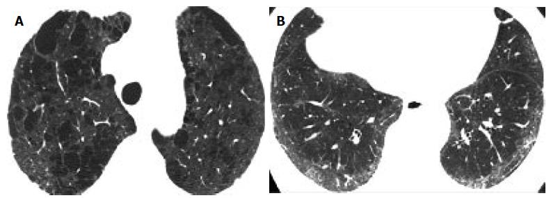
Figure 1 Paraseptal emphysema and usual interstitial pneumonia-pattern of pulmonary fibrosis.
A: High resolution computed tomography (HRCT) at the level of the upper lobes shows paraseptal emphysema associated with subpleural reticular pattern and minimal ground glass opacity; B: HRCT of the same patient as in A, at the level of the lower lobes reveals extensive coarse reticular pattern characterized by traction bronchiectasis associated with ground glass opacity and subpleural honeycombing. The HRCT pattern is consistent more with usual interstitial pneumonia.
Figure 2 Centrilobular emphysema and nonspecific interstitial pneumonia-pattern of pulmonary fibrosis.
A: High resolution computed tomography (HRCT) at the level of the upper lobes shows extensive predominantly centrilobular emphysema; B: HRCT of the same patient as in A, at the level of the lower lobes shows subpleural fine fibrosis characterized mainly by ground glass opacity, reticular pattern non-otherwise specified and absence of honeycombing. The HRCT pattern is consistent more with nonspecific interstitial pneumonia.
- Citation: Oikonomou A, Mintzopoulou P, Tzouvelekis A, Zezos P, Zacharis G, Koutsopoulos A, Bouros D, Prassopoulos P. Pulmonary fibrosis and emphysema: Is the emphysema type associated with the pattern of fibrosis? World J Radiol 2015; 7(9): 294-305
- URL: https://www.wjgnet.com/1949-8470/full/v7/i9/294.htm
- DOI: https://dx.doi.org/10.4329/wjr.v7.i9.294


