Copyright
©The Author(s) 2015.
World J Radiol. Sep 28, 2015; 7(9): 253-265
Published online Sep 28, 2015. doi: 10.4329/wjr.v7.i9.253
Published online Sep 28, 2015. doi: 10.4329/wjr.v7.i9.253
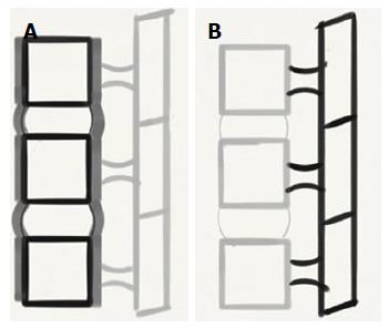
Figure 1 Functional anatomy of the spine (A) and schematic diagrams of the spine (B) depicting the 2 columns in the spine.
Anterior portion of the functional unit (A) consist of two aligned vertebral bodies, the intervertebral disc, the anterior and posterior longitudinal ligaments. Posterior portion (B) consists of the vertebral arches, facet joints, posterior elements and posterior ligamentous complex.
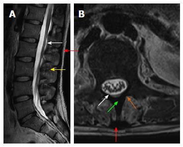
Figure 2 Magnetic resonance anatomy of the spine.
T2 weighted sagittal magnetic resonance (MR) image (A) and T2 weighted axial MR image (B) show posterior ligamentous complex with ligamentumflavum (white arrow), interspinous ligament (yellow arrow) supraspinous ligament (red arrow), spinous process (green arrow) and lamina (orange arrow).
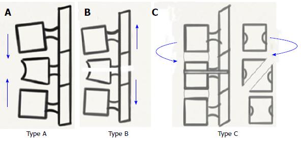
Figure 3 Biomechanics of spinal injury - AO classification.
A-C: Schematic diagrams depicting compression (type A), distraction (type B) and Rotation (type C) injuries.
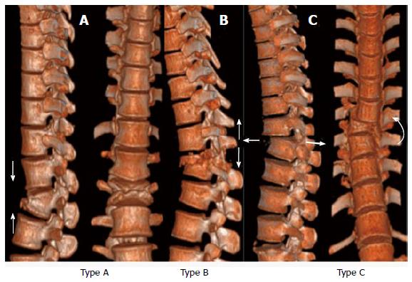
Figure 4 Biomechanics of spinal injury - AO classification.
A-C: Computed tomography Volume rendered images depict compression (type A), Distraction (type B) and Rotation (type C) type injuries.
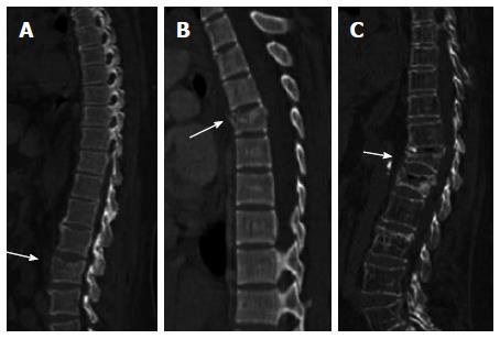
Figure 5 Type A1 compression injuries.
Sagittal computed tomographyimages show (A) end plate impaction (A1.1), (B) wedge impaction (A1.2), and (C) corpus collapse (A1.3).
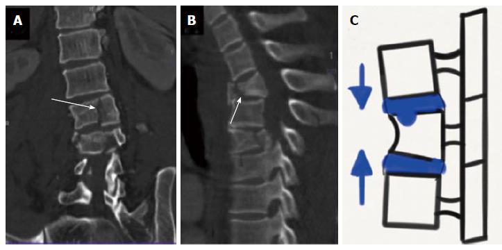
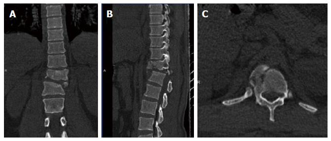
Figure 6 Type A2 compression injuries.
Coronal computed tomography (CT) image (A), sagittal CT image (B) and schematic diagram (C) show (A) sagittal split (A2.1), (B) coronal split (A 2.2) and (C) pincer type injury (A 2.3).
Figure 7 Type A3 compression injuries-burst fractures.
Sagittal computed tomography (CT) image (A), sagittal and coronal CT images (B), sagittal and axial CT images (C) show (A) incomplete burst (A 3.1) (B) burst-split (A 3.2) and (C) retropulsion of fracture fragments suggesting a complete burst injury (A 3.3).
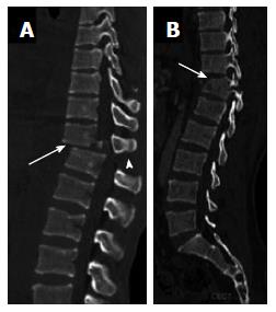
Figure 8 Type B1 flexion distraction injury.
Sagittal computed tomography images show disruption through the posterior ligamentous complex (arrowhead) with either anterior disruption through the disc (arrow) constituting B1.1 injury (A) or a type A fracture anteriorly (arrow) constituting type B1.2 injury (B).
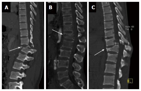
Figure 9 Type B2 flexion distraction injury.
Sagittal computed tomography (CT) image (A), sagittal and coronal CT images (B) and sagittal and axial CT images (C) show transverse bicolumn fracture (B 2.1) [arrow in (A)], flexion-spondylolysis (B 2.2) [arrow in (B)] and (C) flexion- distraction with type A fracture (B 2.3) (arrow).
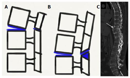
Figure 10 Types of hyperextension injuries (type B3).
Schematic diagrams (A, B) and sagittal CT image (C) showing hyperextension with anterior subluxation (B3.1) (A), hyperextension with spondylolysis and anterior displacement (B3.2) (B) and hyperextension with posterior dislocation (B3.3) [arrow in (C)].
Figure 11 Type C1 rotational injuries.
Coronal computed tomography (CT) reformat (A), sagittal CT reformat (B) and axial CT images (C) show C1 injury where rotation is superimposed on a type A injury.
Figure 12 Type C2 rotational injuries.
Coronal computed tomography (CT) image (A) and sagittal CT images (B) show C2 injury where rotation is superimposed on type B injury.
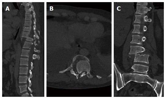
Figure 13 Type C3 rotational injury.
Sagittal CT image (A), axial computed tomography (CT) image (B) and Coronal CT image (C) show shear slice fracture [C3.1 in (A) and (B)] and shear oblique fracture [C3.2 in (C)].
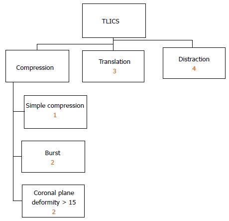
Figure 14 Thoracolumbar injury classification and severity score classification showing points allotted to each injury morphology.
TLICS: Thoracolumbar injury classification and severity score.
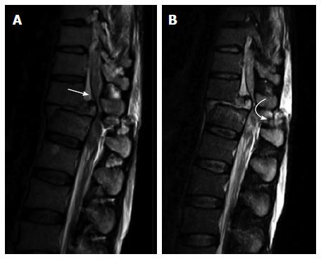
Figure 15 Magnetic resonance imaging in thoraco-lumbar injuries.
T2 weighted sagittal magnetic resonance images showing cord compression (arrow in (A)) and disruption of Posterior Ligamentous Complex [curved arrow in (B)].
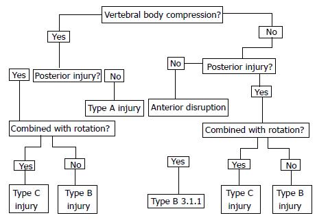
Figure 16 Simplified algorithms showing morphological classification of spinal fractures.
- Citation: Gamanagatti S, Rathinam D, Rangarajan K, Kumar A, Farooque K, Sharma V. Imaging evaluation of traumatic thoracolumbar spine injuries: Radiological review. World J Radiol 2015; 7(9): 253-265
- URL: https://www.wjgnet.com/1949-8470/full/v7/i9/253.htm
- DOI: https://dx.doi.org/10.4329/wjr.v7.i9.253


