Copyright
©The Author(s) 2015.
World J Radiol. Jun 28, 2015; 7(6): 110-127
Published online Jun 28, 2015. doi: 10.4329/wjr.v7.i6.110
Published online Jun 28, 2015. doi: 10.4329/wjr.v7.i6.110
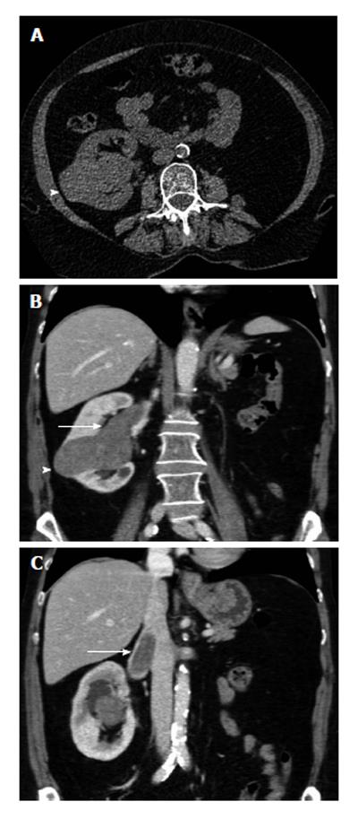
Figure 1 The 65-year-old woman with papillary renal cell carcinoma of the right kidney and tumoral invasion of the ipsilateral renal vein and the inferior vena cava (stage T3b, grade 2).
The patient had left radical nephrectomy years ago for renal cell carcinoma. A: Transverse unenhanced computed tomography (CT) image shows a lobular right renal mass (arrowhead), located in the interlobar region. The mass is relatively homogeneous, slightly hyperdense (CT density: 40 HU), when compared to the normal renal parenchyma; B and C: Contrast-enhanced coronal multiplanar reformations during the corticomedullary phase depict right renal tumor, with moderate, homogeneous enhancement (arrowhead, mean CT density: 65 HU). Venous tumour thrombus is diagnosed as a filling defect within right renal vein and the infrahepatic part of the inferior vena cana (arrow). Neoplastic thrombus is seen extending directly from the neoplasm, enhancing with a similar pattern with primary malignancy.
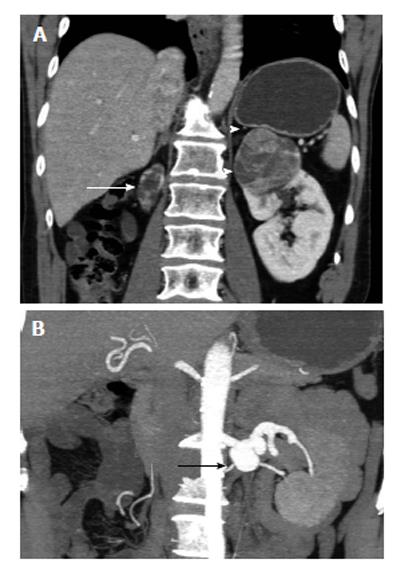
Figure 2 The 70-year-old man with clear cell renal cell carcinoma of the left solitary functioning kidney (stage T1b, grade 2).
A: Post-contrast enhanced coronal multiplanar reformation during the corticomedullary phase depicts left upper pole renal mass, strongly and heterogeneously enhancing, after contrast material administration. A thin hyperdense rim (arrowheads) is detected around the tumor, proved to correspond to fibrous pseudocapsule on pathology. Atrophic right kidney (arrow); B: Coronal 3D-reconstruction during the same phase, using maximum intensity projection algorithm shows left renal artery aneurysm (arrow).
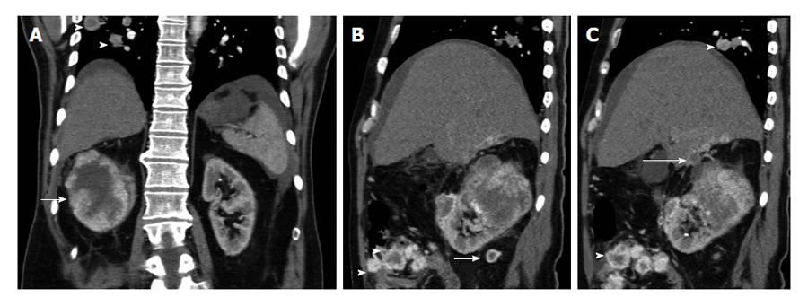
Figure 3 The 52-year-old man with advanced-stage clear cell renal cell carcinoma of the right kidney.
Contrast-enhanced (A) coronal and (B and C) sagittal reformations during the corticomedullar phase show a large right renal tumor (arrow), strongly and inhomogeneously enhancing. Central hypodense parts within malignancy corresponded to areas of necrosis on histology. There is perinephric stranding and contrast-enhancing nodules in the perinephric fat (long arrow, B), a finding strongly suggestive for perinephric fat invasion. The tumor is seen extending and invading the undersurface of the liver (long arrow, C). Lung metastases are detected (arrowheads, A and C). There is also a small amount of ascites and nodular peritoneal masses (arrowheads, B), with heterogeneous enhancement, identical to that of the primary neoplasm, findings suggestive of peritoneal metastases. Peritoneal metastases from renal cell carcinoma (RCC) are extremely rare. Neoplastic invasion of the peritoneum by RCC may occur either, by contiguous spread of renal tumor through the renal capsule, the anterior renal fascia and the posterior parietal peritoneum, or via tumoral emboli.
Figure 4 The 62-year-old man with clear cell renal cell carcinoma of the left kidney (stage T1a, grade II).
A: Transverse plain computed tomography image barely depicts lower pole left kidney mass (arrow), slightly hyperdense. This finding was appreciated after studying the post-contrast enhanced images; B: Coronal reformations during the corticomedullary; C: The nephrographic phase. The tumor (arrow) is seen enhancing strongly and heterogeneously during the corticomedullary phase, a finding strongly suggestive for the diagnosis of renal cell carcinoma (RCC). Hypervascular RCCs as in this case, may enhance to the same degree as the renal cortex and may be mistaken for normal renal parenchyma at the corticomedullary phase. The neoplasm is clearly delineated in the nephrographic phase, detected mainly hypodense, when compared to the contrast-enhancing normal renal parenchyma.
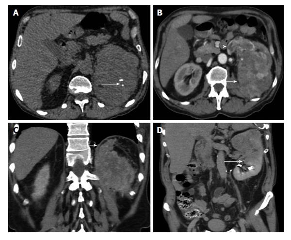
Figure 5 The 62-year-old man with clear cell renal cell carcinoma of the left kidney (stage T3a, grade 3).
A: Transverse unenhanced computed tomography (CT) image shows large heterogenous left renal mass, with small areas of calcifications (long arrow); B: Transverse multiplanar reformation (MPR) during the corticomedullary phase demonstrates left renal malignancy (arrow), inhomogeneously enhancing. The left renal vein is dilated and enhances heterogeneously (arrowhead) due to neoplastic invasion. VTT enhances with a same pattern as renal cell carcinoma; C: Coronal reformations during the corticomedullary phase depicts tumor ill-defined margins and extension into the perinephric fat tissue (arrow). Thickening of the diaphragms of the perinephric space is also seen; D: Coronal MPR during the excretory phase shows nonvisualization of the upper calyces and invasion of the middle calyceal group (arrow), a finding strongly suggestive of invasion of renal sinus fat. CT findings were confirmed both surgically and pathologically.
Figure 6 The 74-year-old man with synchronous bilateral renal cell carcinomas of clear cell type (stage T1).
Bilateral synchronous renal cell carcinomas (RCCs) are uncommon, reported in less than 2% of patients with RCCs. The patient had left radical nephrectomy and right partial nephrectomy. Sagittal multiplanar reformations (MPR) (A) and coronal 3D reformation with volume rendering technique (B) during the corticomedullary phase depict a sharly-demarcated tumor in the lower pole of the left kidney (arrow, A), strongly and heterogeneously enhancing. Sagittal (C) MPR and (D) 3D reformation with the same algorithm depict a second, smaller tumor in the upper pole of the right kidney, with a similar pattern of contrast enhancement. Preoperative information obtained with computed tomography examination enabled conservative surgery for the right renal malignancy.
Figure 7 The 60-year-old woman with clear cell renal cell carcinoma of the left kidney (stage T1a, grade II).
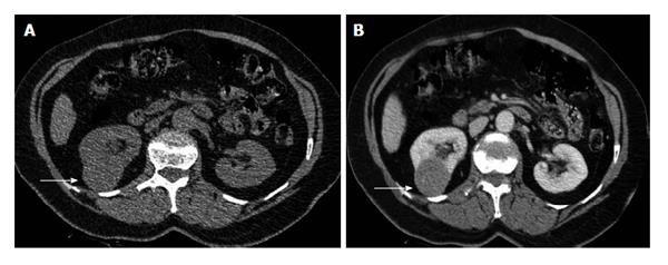
A: Transverse plain computed tomography (CT) image depicts lower pole right renal mass as a focal bulging of the renal contour (arrow), mainly isodense (CT density: 35 HU) to the renal parenchyma; B: Axial multiplanar reformation during the corticomedullary phase clearly shows renal malignancy (arrow) moderately and heterogeneously enhancing (mean CT density: 60 HU). Heterogeneous contrast enhancement on imaging should always suggest renal malignancy preoperatively.
Figure 8 The 75-year-old man with clear cell renal cell carcinoma of the right kidney, invading the liver.
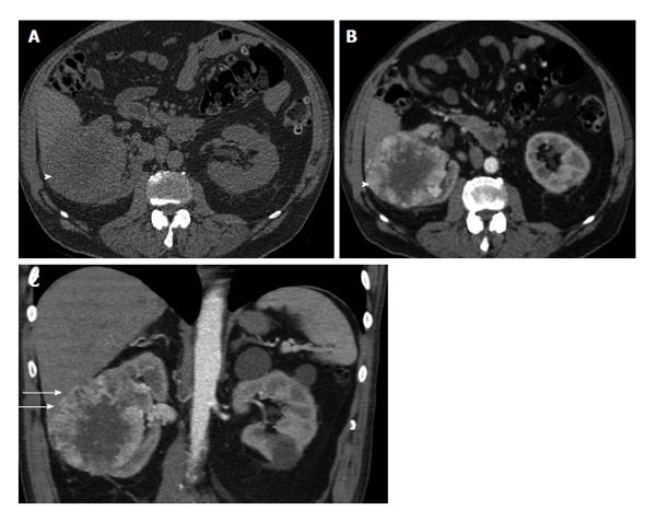
A: Axial plain image shows right heterogeneous right renal tumor (arrowhead); B: Transverse reformation during the corticomedullary phase depicts strong, heterogeneous mass enhancement. The tumor (arrowhead) enhances mainly in the periphery, with a mean computed tomography density of 110 HU (compared to that of 40 HU on the unenhanced images), a finding more compatible with the diagnosis of renal cell carcinoma of the clear cell variety. Central non-enhancing areas corresponded to areas of necrosis on pathology; C: Coronal reformation during the same phase shows renal tumor invading the liver (small arrows), a finding confirmed both on surgery and histopathology.
Figure 9 The 31-year-old man with chromophobe renal cell carcinoma of the left kidney (stage T1b, grade II).
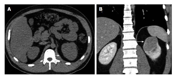
A: Axial plain image barely depicts upper pole left renal mass mainly isodense, with a slight bulging of the renal contour (arrow); B: Coronal reformation during the nephrographic phase clearly depicts left renal tumor (arrow). The neoplasm enhances moderately and homogeneously [computed tomography (CT) density: 70 HU, when compared to the CT density of 35 HU on unenhanced images]. A thin hyperdense rim surrounds renal malignancy, proved to correspond to fibrous pseudocapsule histologically.
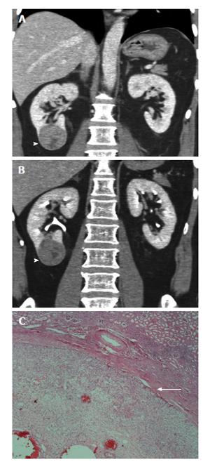
Figure 10 The 50-year-old man with clear cell-chromophobe renal cell carcinoma of right kidney (grade 3, pT1b).
A: Coronal reformatted images in corticomedullary; B: Nephrographic phases depict hyperdense rim (arrowhead) surrounding the tumor; C: Histologic section (H and E, × 400) shows fibrous pseudocapsule (arrow) between tumor and adjacent normal renal parenchyma.
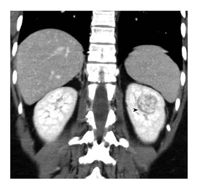
Figure 11 The 44-year-old woman with clear cell renal cell carcinoma of left kidney (grade 2, pT1a).
Computed tomography image demonstrates hypodense rim (arrowhead) around neoplasm detected only on coronal reformations during portal phase. The presence of pseudocapsule was confirmed on histology.
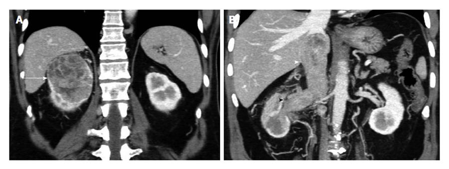
Figure 12 The 64-year-old man with clear cell renal cell carcinoma of the right kidney, invading the renal vein and the inferior vena cava (stage T3b, grade 3).
A: Coronal multiplanar reformations during the corticomedullary phase depicts large, inhomegeneously enhancing right renal tumor (arrow); B: Coronal 3D-display with maximum intensity projection technique during the same phase shows neoplastic thrombus invading left renal vein and the inferior vena cava (arrowheads). Coronal reformations clearly show venous invasion extending below the level of the diaphragm. Perinephric stranding and abnormal vessels are detected in the ipsilateral perinephric space, although pathology was negative for perinephric fat invasion.
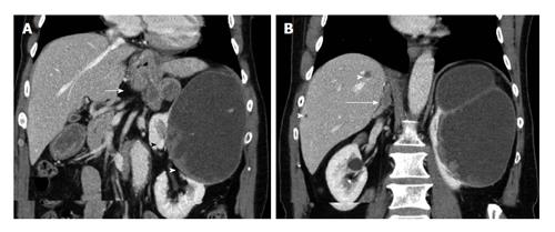
Figure 13 The 70-year-old man with advanced-stage papillary renal cell carcinoma of the left kidney.
Coronal multiplanar reformations during the nephrographic phase show large, mainly cystic left renal mass, with solid contrast-enhancing components (arrowheads). Enlarged retroperitoneal LNs, inhomogeneously enhancing (arrow, A) are detected, compatible with metastatic lymphadenopathy. Liver (arrowhead, B) and right adrenal (long arrow, B) metastases are also seen. All metastatic deposits have a similar pattern of enhancement.
Figure 14 The 81-year-old woman with advanced-stage clear cell renal cell carcinoma of the left kidney.
A: Transverse multiplanar reformation during the nephrographic phase shows large, inhomogeneously enhancing left renal malignancy (arrow), invading the ipsilateral renal vein (arrowhead); B: Contrast-enhanced computed tomography image of the thorax demonstrates enlarged mediastinal lymph nodes (arrow), with heterogeneous enhancement, suggestive for metastatic invasion.
- Citation: Tsili AC, Argyropoulou MI. Advances of multidetector computed tomography in the characterization and staging of renal cell carcinoma. World J Radiol 2015; 7(6): 110-127
- URL: https://www.wjgnet.com/1949-8470/full/v7/i6/110.htm
- DOI: https://dx.doi.org/10.4329/wjr.v7.i6.110


