Copyright
©The Author(s) 2015.
World J Radiol. Oct 28, 2015; 7(10): 329-335
Published online Oct 28, 2015. doi: 10.4329/wjr.v7.i10.329
Published online Oct 28, 2015. doi: 10.4329/wjr.v7.i10.329
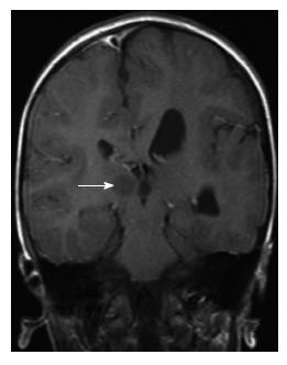
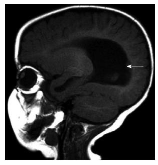
Figure 1 A 10-year-old boy with hemimegalencephaly.
Coronal T1-weighted magnetic resonance image shows hypertrophy of the left cerebral hemisphere, mild enlargement of the left lateral ventricle, hamartoma (white arrow) in postero-medial area of the right thalamus.
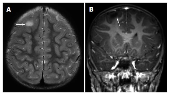
Figure 2 Focal cortical dysplasia type IIb of the left frontal cortex in a 3-year-old girl.
Axial T2-weighted (A) and coronal T1-weighted (B) magnetic resonance images reveal a slight increase of white matter signal on T2-weighted image, significant blurring on T1-weighted image in gray matter-white matter junction (arrow).
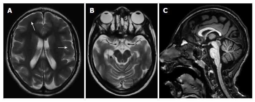
Figure 3 An 8-year-old girl with microlissencephaly.
Axial T2-weighted images (A, B) show reduction in the number and depth of the sulcus, thickened cortex (white arrows) with choroidal fissure cyst (black arrow). Thinning of posterior parts of the corpus callosum is also seen on sagittal three-dimensional (3D) T1-weighted image (C).
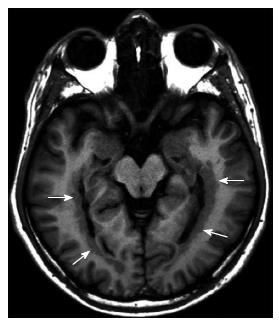
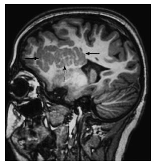
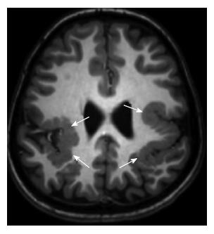
Figure 4 An 18-year-old boy with subependymal heterotopia.
Axial inversion recovery T1-weighted image shows gray matter nodules located adjacent to bilateral periventricular temporal horn region (arrows).
Figure 5 A 7-mo girl with subependymal heterotopia.
Sagittal T1-weighted image shows gray matter nodule located in the periventricular region that protrudes into the ventricular wall (arrow).
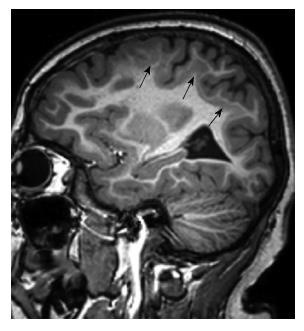
Figure 6 A 12-year-old boy with subcortical heterotopia.
Sagittal 3D T1-weighted image shows large nodular form subcortical heterotopia (arrows) extends from the ventricle into the white matter in frontotemporal region.
Figure 7 An 8-year-old girl with subcortical heterotopia.
Sagittal 3D T1-weighted image shows band heterotopia (double cortex) located between the ventricular wall and the cerebral cortex (arrows).
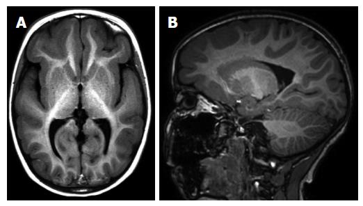
Figure 8 A 5-year-old girl with incomplete lissencephaly.
Axial T1-weighted (A) and sagittal 3D T1-weighted (B) magnetic resonance images show pachygyria in frontotemporal region.
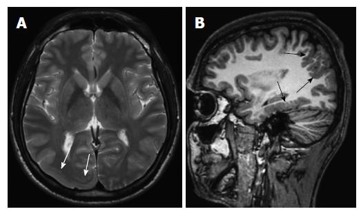
Figure 9 An 18-year-old boy with cobblestone lissencephaly.
Axial T2-weighted image (A) shows incomplete lissencephaly in right occipital region (white arrows). Sagittal 3D T1-weighted image (B) reveals multiple areas of shallow microgyri (black arrows) in the parietal and temporal lobes.
Figure 10 A 7-year-old boy with polymicrogyria.
Axial 3D T1-weighted image shows bilateral perisylvian polymicrogyria (arrows).
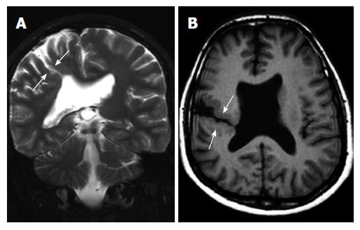
Figure 11 A 12-year-old boy with schizencephaly.
T2-weighted coronal (A) and T1-weighted axial (B) images show oblique gray matter lined holohemispheric cleft (arrows) extending into the lateral ventricle that suggest open lip type schizencephaly with agenesis of septum pellucidum.
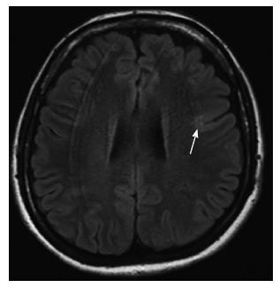
Figure 12 An 18-year-old girl with focal cortical dysplasia type I.
Axial fluid attenuated inversion recovery image shows blurring at gray-white matter junction with normal gray matter signal in the posterior of the left frontal lobe (arrows).
- Citation: Battal B, Ince S, Akgun V, Kocaoglu M, Ozcan E, Tasar M. Malformations of cortical development: 3T magnetic resonance imaging features. World J Radiol 2015; 7(10): 329-335
- URL: https://www.wjgnet.com/1949-8470/full/v7/i10/329.htm
- DOI: https://dx.doi.org/10.4329/wjr.v7.i10.329


