Copyright
©The Author(s) 2015.
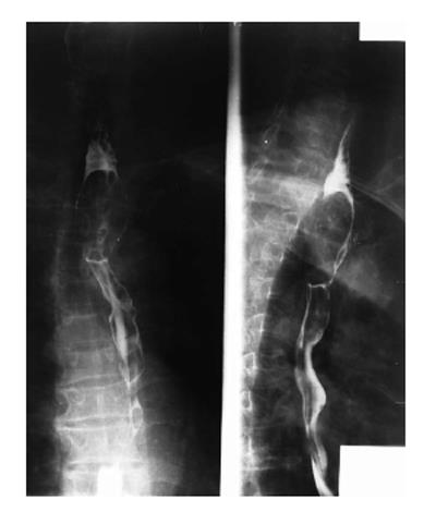
Figure 1 A 60-year-old male patient with squamous cell carcinoma in the first third section of esophagus.
Polypoid-type lesions, presented as a filling defect with luminal narrowing and without ulceration.
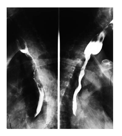
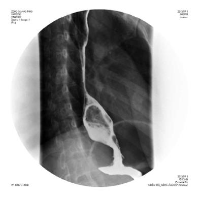
Figure 2 A 60-year-old male with squamous cell carcinoma.
On barium esophagography, infiltration in the upper third of esophageal carcinoma, with irregular luminal narrowing, mucosal destruction, dilatation and abrupt proximal borders. Prestenotic dilatation is also present.
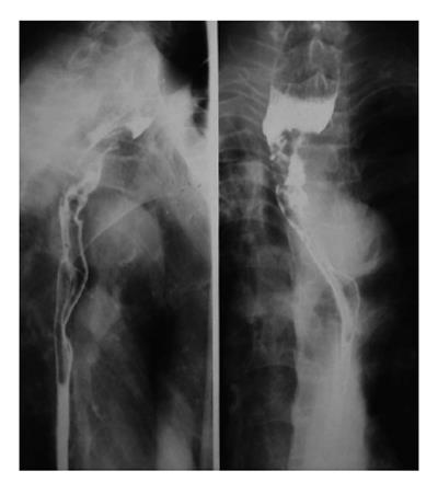
Figure 3 A 59-year-old male with advanced esophageal carcinoma.
Asymmetric stenosis of the first third esophagus, with central, large irregular ulceration, surrounded by a radiolucent rim of neoplasmatic infiltration.
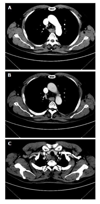
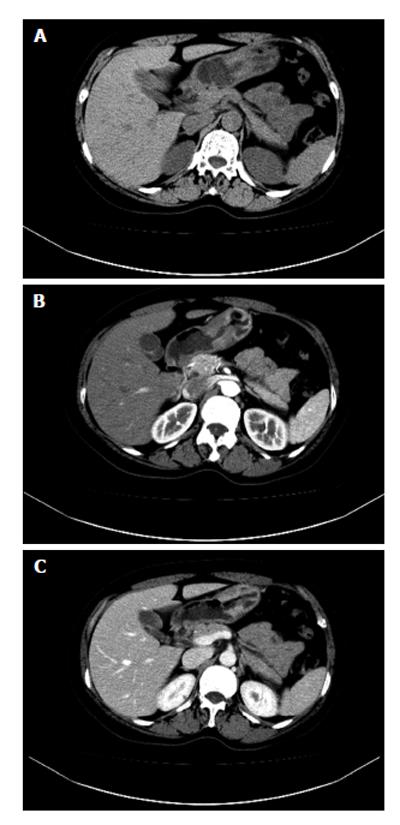
Figure 4 A 61-year-old male with squamous cell carcinoma in the first third section of esophagus.
At computed tomography, esophageal cancer shows a soft tissue mass, with irregular luminal narrowing. The located wall thickening was asymmetric. The lesion shows moderate arterial phase enhancement (A) and slight venous phase enhancement (B). The enlarged lymph node metastasis is shown in the upper right paratracheal (C).
Figure 5 A 29-year-old female with leiomyoma.
A large exophytic submucosal mass with a sharply defined, smooth filling defect is shown in the distal esophagus.
Figure 6 A 29-year-old female with leiomyoma.
Esophageal leiomyoma shows sharply marginated homogeneous mass in the lower third esophagus. These tumors show isoattenuating of slight hypoattenuating to muscle at non-enhanced computed tomography (A), moderate arterial (B) and venous (C) phase enhancement.
Figure 7 A 56-year-old female patient with early gastric carcinoma.
Computed tomography scans showing thickening wall and narrowing antrum of stomach (A), moderate arterial (B) and venous (C) phase enhancement.
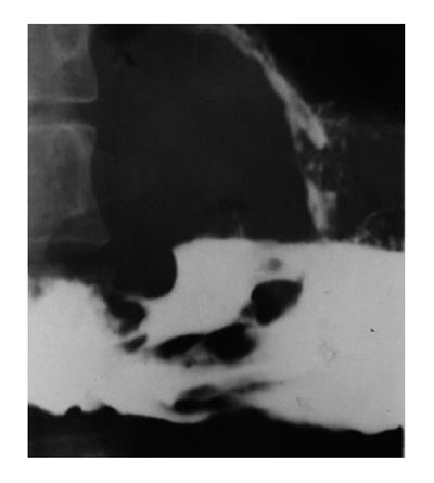
Figure 8 A 62-year-old male with ulcerated gastric carcinoma: a huge ulcerated mass is seen in the gastric antrum.
The margins of the neoplasm’s tissue surrounding the irregular ulcer are also seen.
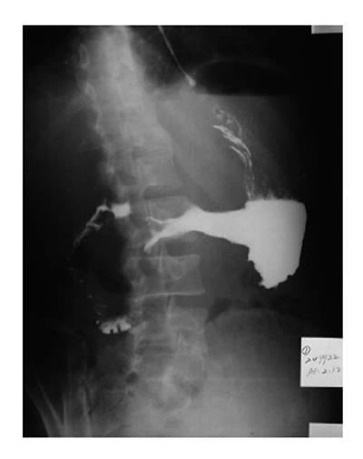
Figure 9 A 56-year-old male.
Diffuse, marked gastric narrowing, with irregular contour and thickened spiculated folds due to primary scirrhous carcinoma.
Figure 10 A 60-year-old female patient with mucosa-associated lymphoid tissue in gastric antrum.
The tumor reveals as segmental, smooth homogeneous wall thickening on non-contrast computed tomography (A), minimal enhancement on arterial (B) and venous (C) phase enhancement.
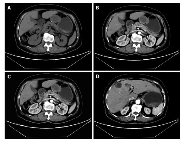
Figure 11 A 76-year-old female with gastrointestinal stromal tumors.
Axial plain (A) shows an round, exophytic soft tissue mass, appearing as a dominant mass extrinsic to the wall of the stomach, contrast-enhanced computed tomography arterial (B) and venous phase (C) scan shows slightly enhancing, occasionally with coarse calcification. Liver metastases were shown (D).
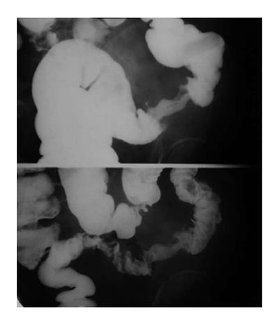
Figure 12 A 59-year-old female patient with sigmoid carcinoma.
An annular stenosis of the sigmoid colon, with an irregular mucosal surface with contour deformity.
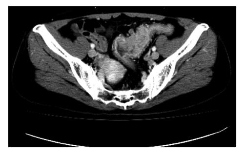
Figure 13 Sigmoid colon cancer in a 59-year-old female.
Contrast-enhanced spiral computed tomography scan shows luminal narrowing and marked wall thickening. There is adjacent stranding of the serosa and mesenteric fat, a small lymph node anterior to the irregular thickening wall.
- Citation: Li YZ, Wu PH. Conventional radiological strategy of common gastrointestinal neoplasms. World J Radiol 2015; 7(1): 7-16
- URL: https://www.wjgnet.com/1949-8470/full/v7/i1/7.htm
- DOI: https://dx.doi.org/10.4329/wjr.v7.i1.7


