Copyright
©2014 Baishideng Publishing Group Inc.
World J Radiol. Sep 28, 2014; 6(9): 730-736
Published online Sep 28, 2014. doi: 10.4329/wjr.v6.i9.730
Published online Sep 28, 2014. doi: 10.4329/wjr.v6.i9.730
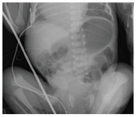
Figure 1 This plain film illustrates an infant with malposition of the small bowel on the right and large bowel on the left suggesting malrotation.
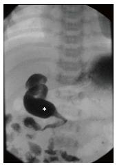
Figure 2 This Upper gastrointestinal demonstrates abnormal position of the duodenal-jejunal junction (white star) to the right of the spine.
Normally the duodenum should sweep across from right to left across the spine.
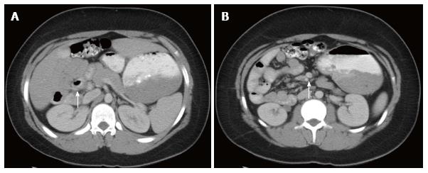
Figure 3 This axial view of an abdominal computed tomography.
A: Illustrates the duodenal-jejunal junction (white arrow) in the right hemi-abdomen suggesting malrotation; B: Illustrates superior mesenteric artery Superior Mesenteric Artery (SMA)/Superior Mesenteric Vein (SMV) inversion (white arrow) with the SMA to the right of the SMV. This inversion suggests malrotation.
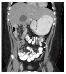
Figure 4 This coronal view of an abdominal computed tomography illustrates the terminal ileum and cecum (white arrows).
Positioning of the cecum in the left hemi-abdomen is suggestive of malrotation.
Figure 5 This Upper gastrointestinal in an infant with heterotaxia demonstrates abnormal positioning of the stomach to the right, the liver near the midline, and the duodenum running left to right.
The duodenal-jejunal junction (white arrow) is seen inferior to the duodenum demonstrating malrotation.
- Citation: Tackett JJ, Muise ED, Cowles RA. Malrotation: Current strategies navigating the radiologic diagnosis of a surgical emergency. World J Radiol 2014; 6(9): 730-736
- URL: https://www.wjgnet.com/1949-8470/full/v6/i9/730.htm
- DOI: https://dx.doi.org/10.4329/wjr.v6.i9.730


