Copyright
©2014 Baishideng Publishing Group Inc.
World J Radiol. Aug 28, 2014; 6(8): 629-635
Published online Aug 28, 2014. doi: 10.4329/wjr.v6.i8.629
Published online Aug 28, 2014. doi: 10.4329/wjr.v6.i8.629
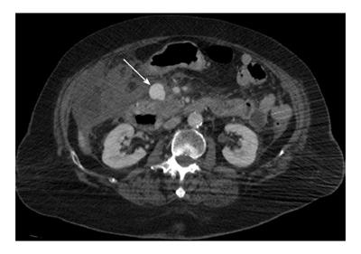
Figure 1 Axial contrast-enhanced computed tomography showing pancreatico-duodenal artery pseudoaneurysm (arrow) before the treatment.
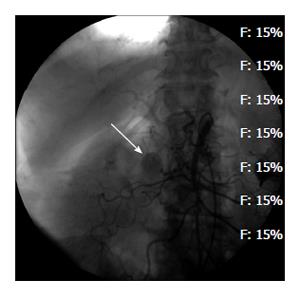
Figure 2 Selective superior mesenteric artery catheterization showing pancreatico-duodenal artery pseudoaneurysm (arrow) but the feeding vessel could not be identified.
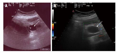
Figure 3 Two-dimensional B-mode ultrasonography (A) and color-Doppler ultrasonography (B) showing pancreatico-duodenal artery pseudoaneurysm (arrows) before and after percutaneous thrombin injection, respectively.
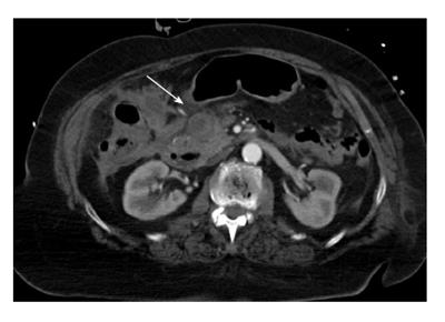
Figure 4 Axial contrast-enhanced computed tomography showing pancreatico-duodenal artery pseudoaneurysm (arrow) 1 wk after the treatment.
- Citation: Barbiero G, Battistel M, Susac A, Miotto D. Percutaneous thrombin embolization of a pancreatico-duodenal artery pseudoaneurysm after failing of the endovascular treatment. World J Radiol 2014; 6(8): 629-635
- URL: https://www.wjgnet.com/1949-8470/full/v6/i8/629.htm
- DOI: https://dx.doi.org/10.4329/wjr.v6.i8.629


