Copyright
©2014 Baishideng Publishing Group Inc.
World J Radiol. Jul 28, 2014; 6(7): 417-423
Published online Jul 28, 2014. doi: 10.4329/wjr.v6.i7.417
Published online Jul 28, 2014. doi: 10.4329/wjr.v6.i7.417
Figure 1 Coronal view of the maxillary molar with multi-detector computed tomography.
The absence of cortication between the root apex and maxillary sinus can be observed.
Figure 2 Root darkening and inferior alveolar canal shape.
A: Darkening of the root is observed in this orthopantomography image. A black band is visible (white arrows) at the root apex site; B: A cross-sectional multi-detector computed tomography image indicates alterations (dumbbell shape) to the inferior alveolar canal shape and a loss of the lingual cortex of the mandibular bone.
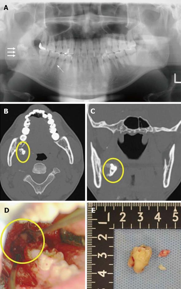
Figure 3 Imaging and surgical removal of a dislocated third mandibular molar (LM3).
A: Orthopantomography showing a dislocated LM3 during surgical removal. The dislocated LM3 is superimposed over the middle part of the mandibular ramus (white arrows); B: Axial multi-detector computed tomography (MDCT) image and C: coronal MDCT image of the dislocated LM3 deviating into the pterygomandibular space (yellow circle); D: Surgical extraction; E: Extracted LM3.
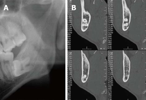
Figure 4 Radiolucency is not observed with orthopantomography.
A: No significant radiolucency is observed in the roots of the third mandibular molar with orthopantomography; B: A cross-sectional multi-detector computed tomography image shows the dumbbell shape of the inferior alveolar canal and loss of the lingual cortex of the mandibular bone.
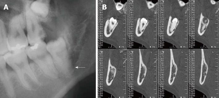
Figure 5 Root darkening and altered third mandibular molar (LM3) root.
A: Darkening of the root observed by orthopantomography. A black band is visible (white arrow) at the root apex site; B: Alteration of the LM3 root (grooved) and loss of the lingual cortex of mandibular bone can be observed on cross-sectional multi-detector computed tomography images.
- Citation: Nakamori K, Tomihara K, Noguchi M. Clinical significance of computed tomography assessment for third molar surgery. World J Radiol 2014; 6(7): 417-423
- URL: https://www.wjgnet.com/1949-8470/full/v6/i7/417.htm
- DOI: https://dx.doi.org/10.4329/wjr.v6.i7.417


