Copyright
©2014 Baishideng Publishing Group Co.
World J Radiol. Apr 28, 2014; 6(4): 125-129
Published online Apr 28, 2014. doi: 10.4329/wjr.v6.i4.125
Published online Apr 28, 2014. doi: 10.4329/wjr.v6.i4.125
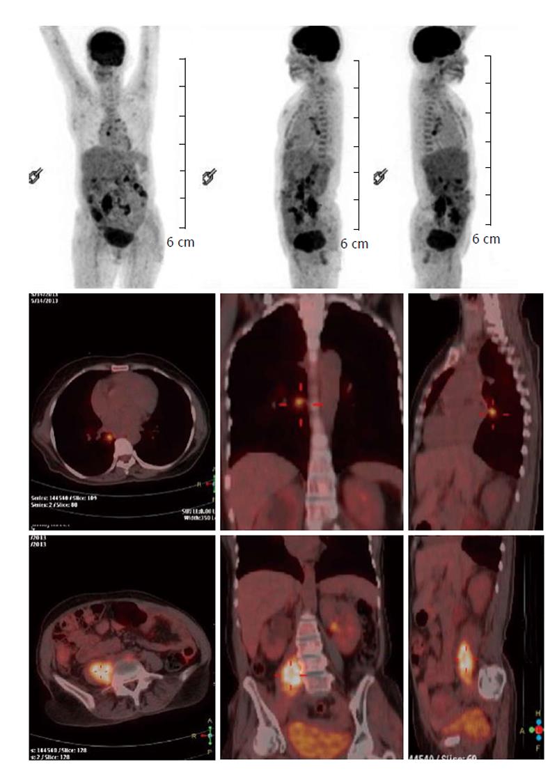
Figure 1 Whole body fluorodeoxyglucose-positron emission tomography/computed tomography.
This first suggested disease involvement of the psoas muscle demonstrating intense FDG uptake (compared with the contralateral muscle). In addition, FDG foci are noted in the prevertebral, left paraaortic, peribronchial and hilar lymph nodes. FDG: Fluorodeoxyglucose.
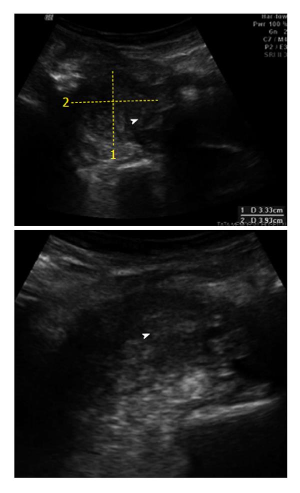
Figure 2 Ultrasound of the abdomen demonstrating a bulky right psoas muscle with heterogenous echotexture.
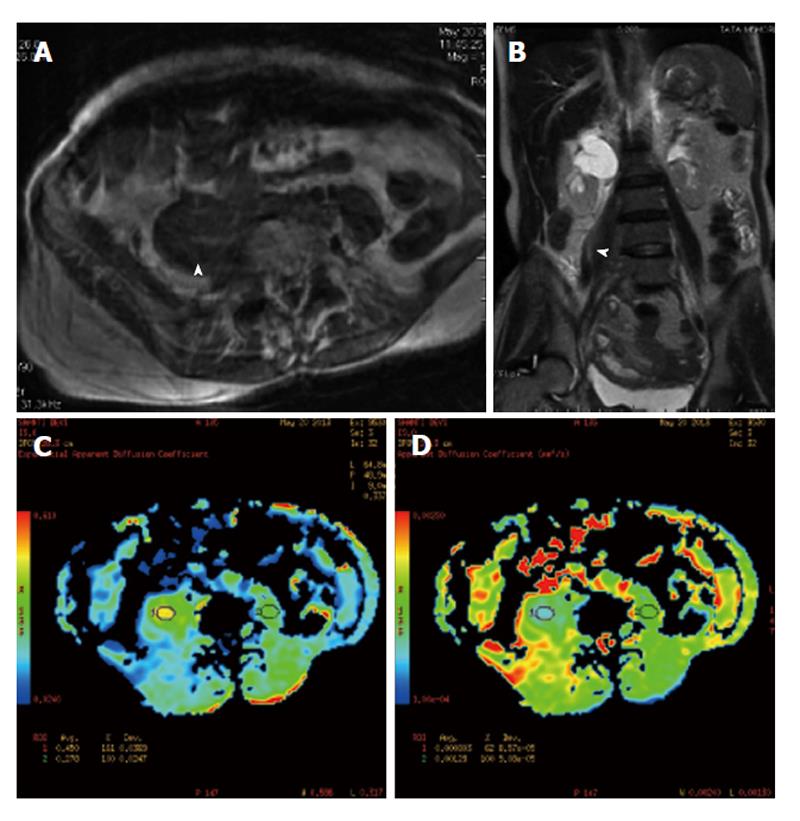
Figure 3 Magnetic resonance imaging.
Axial T1W and coronal T2W equence (A and B) reveals a bulky right psoas muscle (arrowhead) and shows altered signal intensity. On axial diffusion weighted images (C and D) there is e/o restricted diffusion in the involved segment.
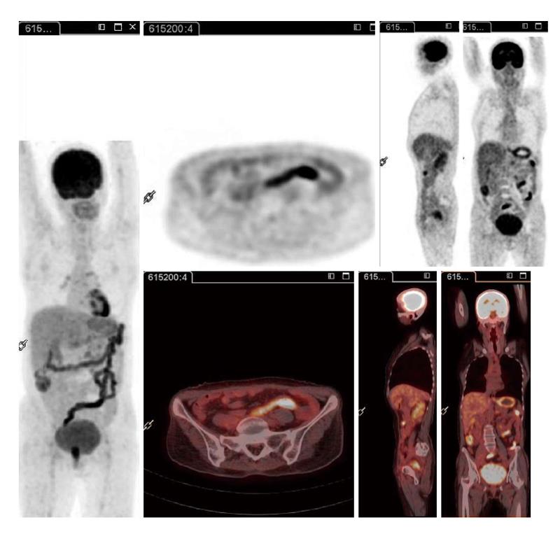
Figure 4 Follow-up fluorodeoxyglucose-positron emission tomography/computed tomography following administration of 3 cycles of systemic chemotherapy and local external radiotherapy demonstrating near-complete resolution of the fluorodeoxyglucose uptake in the psoas muscle as well as other foci.
- Citation: Basu S, Mahajan A. Psoas muscle metastasis from cervical carcinoma: Correlation and comparison of diagnostic features on FDG-PET/CT and diffusion-weighted MRI. World J Radiol 2014; 6(4): 125-129
- URL: https://www.wjgnet.com/1949-8470/full/v6/i4/125.htm
- DOI: https://dx.doi.org/10.4329/wjr.v6.i4.125


