Copyright
©2014 Baishideng Publishing Group Inc.
World J Radiol. Dec 28, 2014; 6(12): 932-936
Published online Dec 28, 2014. doi: 10.4329/wjr.v6.i12.932
Published online Dec 28, 2014. doi: 10.4329/wjr.v6.i12.932
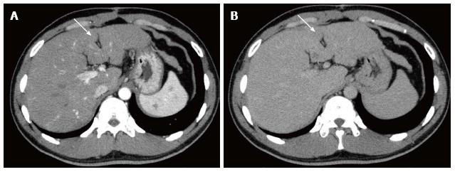
Figure 1 Multidetector-row computed tomography.
A: Arterial phase shows an area of little enhancement (arrow) in segment IV; B: Equilibrium phase shows an area of hypodensity (arrow) as compared with surrounding liver parenchyma.
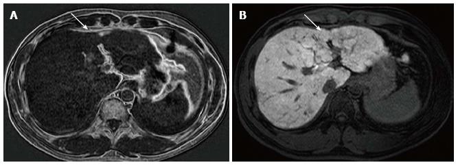
Figure 2 Gadoxetate disodium-enhanced magnetic resonance imaging.
A: Subtraction chemical shift images before contrast enhancement shows high intensity of the lesion (arrow), suggesting the presence of small amount of fat; B: On hepatobiliary phase, an area of diminished uptake of gadoxetate disodium is seen (arrow), which was slightly smaller in size than the fatty area shown on A.
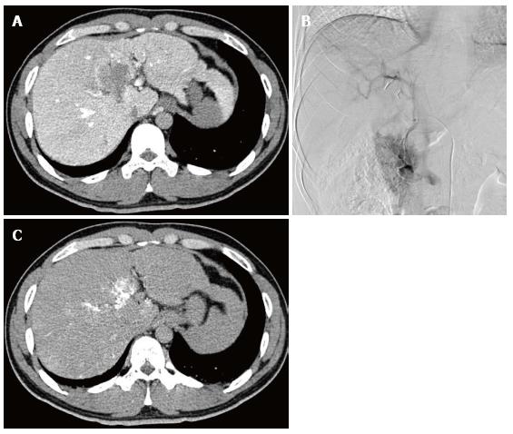
Figure 3 Angiography-assisted computed tomography.
A: Computed tomography (CT) during arterial portography shows a portal venous perfusion defect in S4, which was significantly larger in size than the fatty area shown on CT or magnetic resonance imaging; B: Venous phase of gastroduodenal arteriogram shows an unusual vessel coursing towards the hepatic hilum, representing an aberrant pancreatico-duodenal vein; C: Venous phase of CT during gastroduodenal arteriogram shows that the venous blood from the region of the head of the pancreas directly drains the areas of portal perfusion defect as shown in 3A.
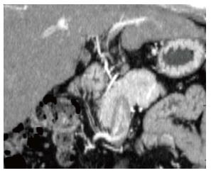
Figure 4 Curved reformation image of the arterial phase of computed tomography reconstructed along the aberrant vessel.
The whole course of the aberrant pancreatico-duodenal vein is well demonstrated. The computed tomography data used here are the ones obtained before cholecystectomy.
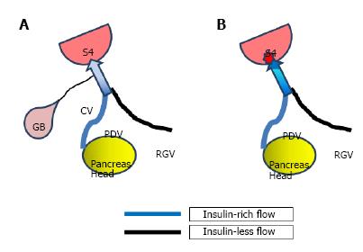
Figure 5 Schematic diagram showing the possible mechanism of the development of focal fatty change in segment IV in the present case.
A: Before cholecystectomy. Insulin-rich flow from the head of the pancreas may be diluted by venous blood that does not contain insulin. S4 of the liver may be normal in appearance, slightly enhanced, or slightly fatty, depending on the amount of blood flow and insulin conveyed to S4; B: After cholecystectomy. Because there is no more diluting blood flow from the gallbladder, insulin-rich flow from the head of the pancreas may directly reach S4 of the liver, where intense focal fatty change may occur. S4: Segment IV; PDV: Pancreaticoduodenal vein; RGV: Right gastric vein.
- Citation: Osame A, Mitsufuji T, Kora S, Yoshimitsu K, Morihara D, Kunimoto H. Focal fatty change in the liver that developed after cholecystectomy. World J Radiol 2014; 6(12): 932-936
- URL: https://www.wjgnet.com/1949-8470/full/v6/i12/932.htm
- DOI: https://dx.doi.org/10.4329/wjr.v6.i12.932


