Copyright
©2014 Baishideng Publishing Group Inc.
World J Radiol. Dec 28, 2014; 6(12): 928-931
Published online Dec 28, 2014. doi: 10.4329/wjr.v6.i12.928
Published online Dec 28, 2014. doi: 10.4329/wjr.v6.i12.928
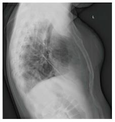
Figure 1 Lateral chest X-ray demonstrates anterior low-density soft tissue mass overlying sternum.
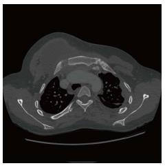
Figure 2 Axial computed tomography demonstrates soft tissue mass anteriorly, centered on the costo-clavicular joint with mediastinal extension.
Note the destructive change at the medial margin of the first and the lateral margin of the sternum.
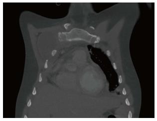
Figure 3 Coronal computed tomography demonstrates some destructive change of the anterior first rib and widening of the first costo-clavicular joint.
- Citation: Patel P, Gray RR. Tuberculous osteomyelitis/arthritis of the first costo-clavicular joint and sternum. World J Radiol 2014; 6(12): 928-931
- URL: https://www.wjgnet.com/1949-8470/full/v6/i12/928.htm
- DOI: https://dx.doi.org/10.4329/wjr.v6.i12.928


