Copyright
©2014 Baishideng Publishing Group Inc.
World J Radiol. Oct 28, 2014; 6(10): 833-839
Published online Oct 28, 2014. doi: 10.4329/wjr.v6.i10.833
Published online Oct 28, 2014. doi: 10.4329/wjr.v6.i10.833
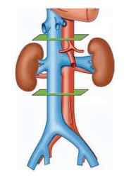
Figure 1 Inferior vena cava studied levels.
The juxtarenal level is the site of the entrance of the renal veins; at 3 cm to the juxtarenal level are the suprarenal (above) and the infrarenal (below) levels, marked in green.
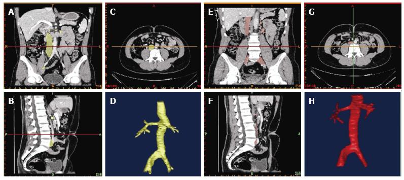
Figure 2 Three-dimensional inferior vena cava reconstruction in neutral breathing and in Valsalva.
The figure shows different perspectives in order to get the different shapes of the vena cava cross-section. The inferior vena cava is marked in yellow during neutral breathing (A-D) and in red during the Valsalva maneuver (E-H). A, E: Coronal cross-section; B, F: Sagittal cross-section; C, G: Axial cross-section; D, H: Reconstruction of the inferior vena cava.
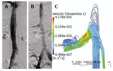
Figure 3 Distribution forces of the contrast medium in the inferior vena cava.
A: Phlebography in neutral breathing; B: Phlebography during the Valsalva maneuver that shows distribution of the contrast medium to renal and iliac veins; C: Computational study of inferior vena cava migration forces using an experimental set up and finite element simulation during the Valsalva maneuver. When the patient is asked to perform a Valsalva maneuver, the inferior vena cava blurs as the contrast is displaced to lower pressure areas, such as iliacs and renal veins.
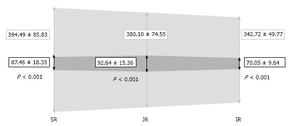
Figure 4 Mean area values in each level and their standard deviation (mm2).
During neutral breathing (in light grey) and during Valsalva (in dark grey); SR: Suprarenal; JR: Juxtarenal; IR: Infrarenal.
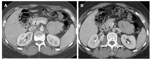
Figure 5 Angio computed tomography.
Images showing the suprarenal inferior vena cava (arrows). A: In neutral breathing; B: In the Valsalva maneuver.
Figure 6 Three-dimensional reconstruction of the inferior vena cava.
Modeling at various perspectives showing the different shapes of the vena cava cross-section in A: Neutral breathing; B: During the Valsalva maneuver.
- Citation: Laborda A, Sierre S, Malvè M, Blas ID, Ioakeim I, Kuo WT, Gregorio MAD. Influence of breathing movements and Valsalva maneuver on vena caval dynamics. World J Radiol 2014; 6(10): 833-839
- URL: https://www.wjgnet.com/1949-8470/full/v6/i10/833.htm
- DOI: https://dx.doi.org/10.4329/wjr.v6.i10.833


