Copyright
©2013 Baishideng Publishing Group Co.
World J Radiol. Sep 28, 2013; 5(9): 349-351
Published online Sep 28, 2013. doi: 10.4329/wjr.v5.i9.349
Published online Sep 28, 2013. doi: 10.4329/wjr.v5.i9.349
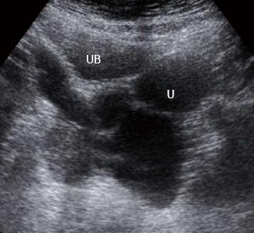
Figure 1 Ultrasound of the pelvis shows a multilocular cystic lesion posterior to the bladder to the left.
U: Uterus; UB: Urinary bladder.
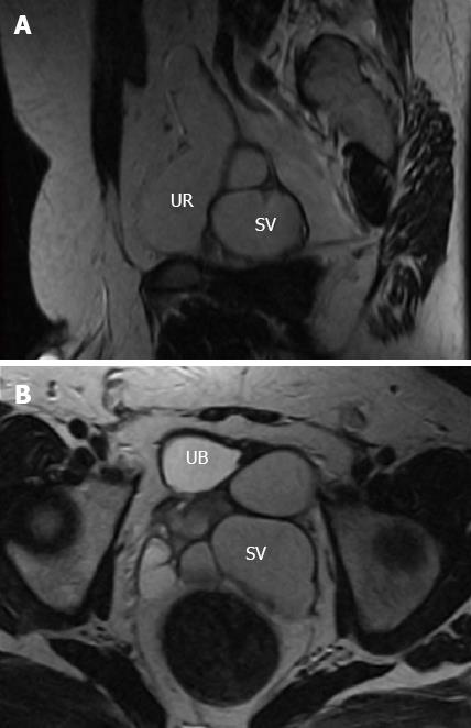
Figure 2 Sagittal and Axial T2w magnetic resonance imaging.
A: The left ureter stump with ectopic insertion into the dilated left seminal vesicle (SV); B: Altered fluid content at the dilated left seminal vesicle with relative low small intestine. UB: Urinary bladder; UR: Ureter.
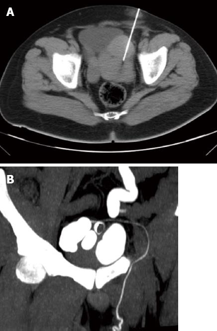
Figure 3 Axial and coronal Computed Tomography.
A: The pelvis with needle punctures of the left seminal vesicle; B: Contrast opacification of the seminal vesicle, vas deferens and left ureter stump.
- Citation: El-Ghar MA, El-Diasty T. Ectopic insertion of the ureter into the seminal vesicle. World J Radiol 2013; 5(9): 349-351
- URL: https://www.wjgnet.com/1949-8470/full/v5/i9/349.htm
- DOI: https://dx.doi.org/10.4329/wjr.v5.i9.349


