Copyright
©2013 Baishideng Publishing Group Co.
World J Radiol. Aug 28, 2013; 5(8): 275-284
Published online Aug 28, 2013. doi: 10.4329/wjr.v5.i8.275
Published online Aug 28, 2013. doi: 10.4329/wjr.v5.i8.275
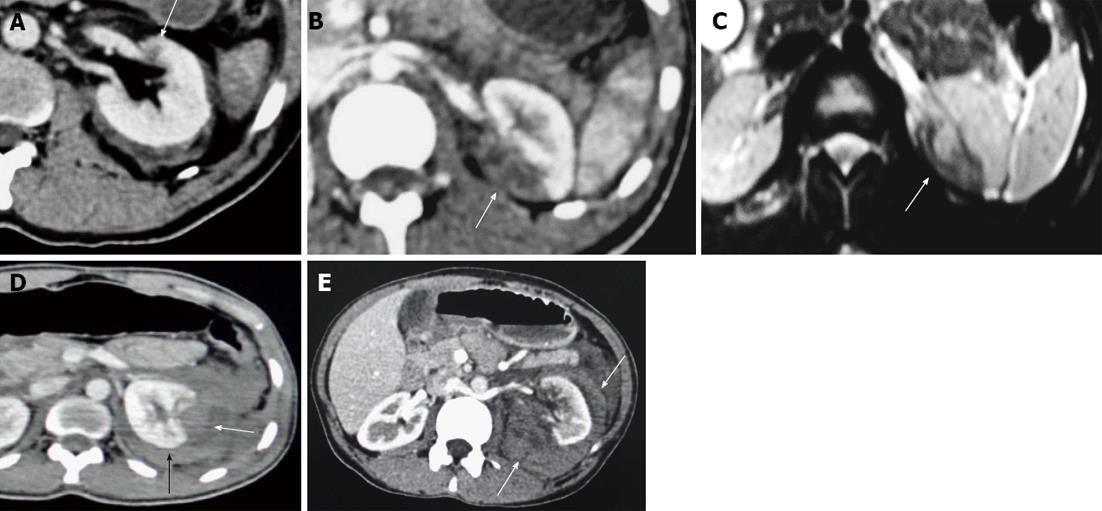
Figure 1 Terminology constellation.
A: Contusion. Axial contrast enhanced computed tomography (CECT) section showing an ill-defined and poorly marginated hypodense area of decreased enhancement (arrow) suggestive of intra-parenchyma contusion; B, C: Infarction; B: Axial CECT; C: Axial T2 weighted magnetic resonance imaging (MRI) image showing infarcts as wedge shaped, sharply marginated hypodense area on computed tomography (arrow) and hypointense area on T2W MRI image (arrow); D: Subcapsular hematoma. Axial CECT section showing hypodense area of blood collection along the renal contour causing flattening or depression of the underlying renal surface (arrow). Also, there is associated perirenal hematoma (black arrow); E: Perirenal hematoma. Axial CECT image showing hypodense area of fluid collection (arrows) around the left kidney confined within the Gerota’s fascia and not producing flattening of the renal contour.
Figure 2 Grade I injury.
A: Axial; B: Coronal contrast enhanced computed tomography (CECT) image of a 40-year-old male patient who had sustained a road traffic accident, shows a small contusion at upper pole of left kidney (Grade I injury). Also seen is a small wedge shaped hypodense area of infarct at lower pole of right kidney; C: Axial CECT image of another patient (25-year-old male with blunt trauma) showing a larger area of contusion at upper pole of right kidney.
Figure 3 Grade II injury.
A: Axial contrast enhanced computed tomography (CECT) section of 24-year-old male patient who had blunt trauma to the abdomen, showing a small superficial irregular and linear hypodense area of laceration (arrow) involving the posterior cortex of left kidney with mild perinephric hematoma. No pelvicalyceal system injury; B: Axial; C: Coronal CECT images of 27-year-old male patient with blunt trauma showing perirenal hypodensity (arrows) surrounding the left kidney and confined within the Gerota’s fascia.
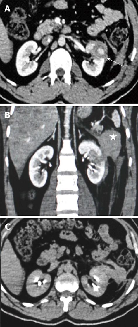
Figure 4 Grade III injury.
Contrast enhanced computed tomography (CECT) images of a 27-year-old male patient who had a stab injury in the left flank. A: Axial section showing a deep laceration (arrow) reaching up to the hilum; B: Coronal section showing the deep laceration at mid pole with disruption of the pararenal fascia and a large hematoma (star) lateral to it; C: Axial delayed image in excretory phase showed no excreted contrast extravasation.
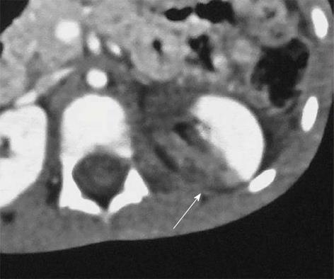
Figure 5 Grade IV injury.
Segmental infarction. Contrast enhanced computed tomography axial section of a 6-year-old child who sustained blunt trauma. Sharply marginated nonenhancing hypodense area involving the posteromedial cortex of left kidney.
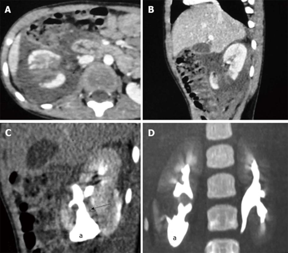
Figure 6 Grade IV injury.
Laceration involving the pelvicalyceal system (PCS). Contrast enhanced computed tomography (CECT) images of a 7-year-old male who had a fall from height with resulting blunt trauma to the abdomen. A: Axial; B: Sagittal section showing deep laceration in lower pole of the right kidney extending up to the renal hilum; C: Sagittal section; D: Thick coronal maximum intensity projection image of the excretory phase shows leakage of excreted opacified urine (arrow) through the laceration suggesting PCS injury with resultant urinoma (a) formation around lower pole of right kidney.
Figure 7 Grade IV injury.
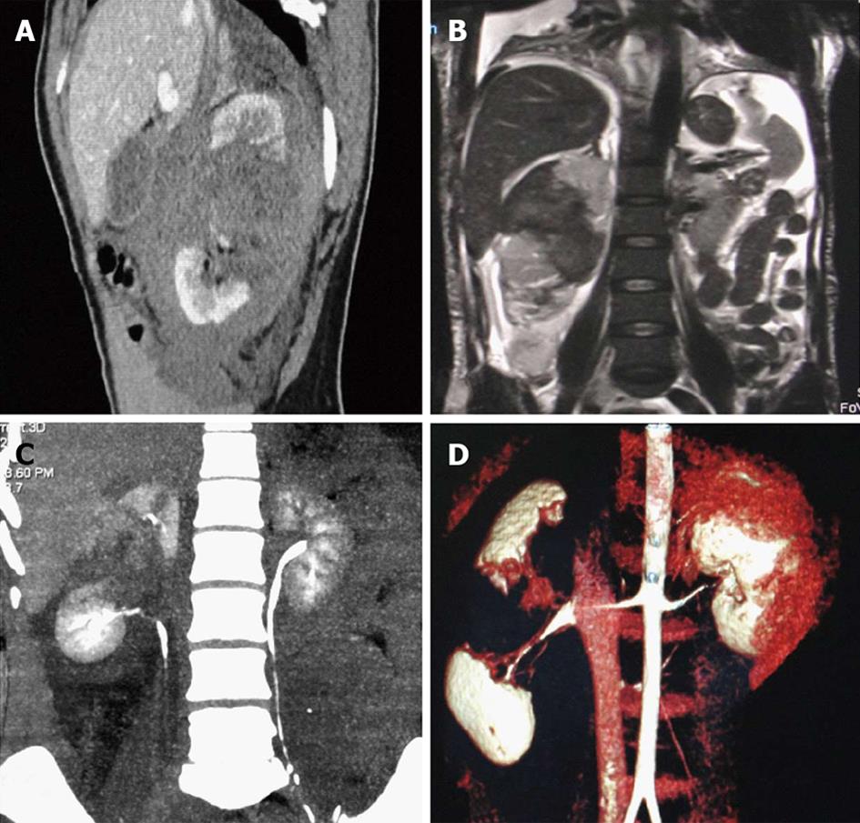
Fractured kidney. Contrast enhanced computed tomography (CECT) and magnetic resonance imaging (MRI) images of a 58-year-old female patient who sustained renal tubular acidosis and had significant hematuria. A: Sagittal CECT; B: Coronal T2 weighted MRI section showing a large laceration with hematoma extending through the mid pole of the right kidney dividing the kidney into two parts and separating the poles wide apart; C: Coronal maximum intensity projection image of the excretory phase did not show any urinary leak with opacification of the ureter; D: Volume rendered reconstructed image showing the fractured kidney through its waist.
Figure 8 Grade V injury.
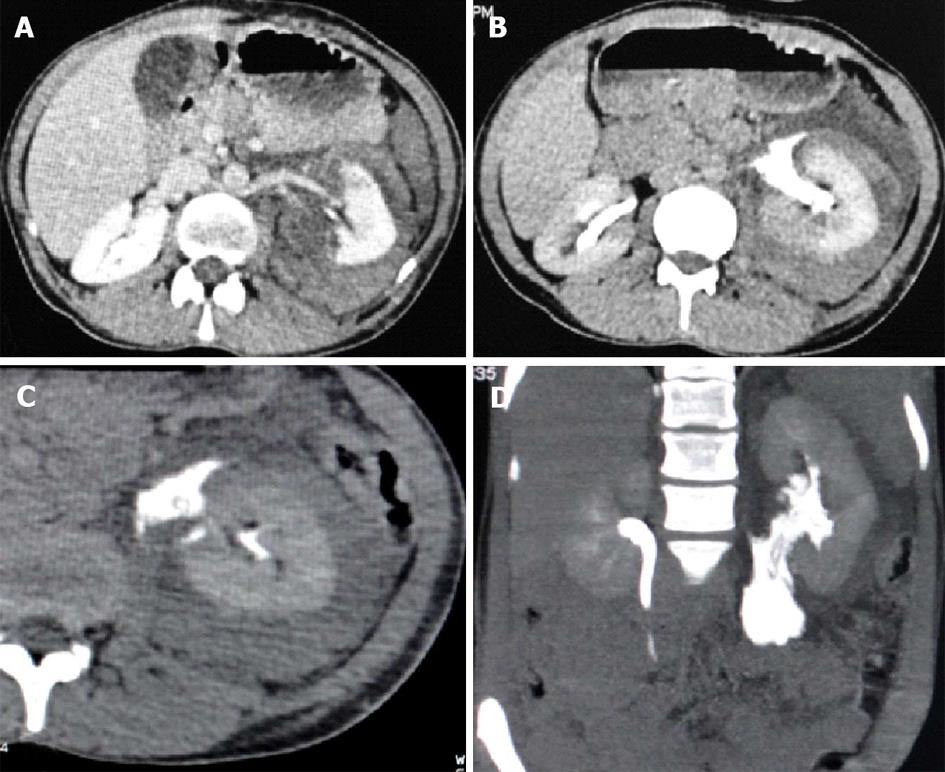
Ureteropelvic junction injury (revised stage Grade IV). Contrast enhanced computed tomography images of a 30-year-old female patient who sustained blunt abdominal trauma. A: Axial section showing deep laceration in posteromedial cortex of left kidney involving the hilum with perinephric and perihilar hematoma; B: Excretory phase axial section showing leak of opacified urine from the pelviureteric junction which tracks along the medial aspect of kidney; C: Axial maximum intensity projection (MIP); D: Coronal MIP image showing the extravasated opacified urine from the renal pelvis and tracking in the retroperitoneum along the course of ureter.
Figure 9 Grade V injury.
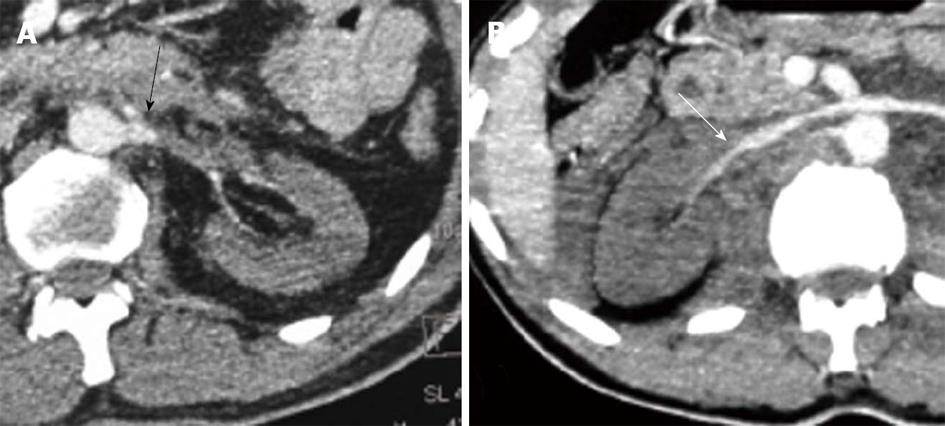
Renal vascular injury. Devascularization following renal artery thrombosis. A: Axial contrast enhanced computed tomography (CECT) section at the level of left renal artery of a 48-year-old male patient who had blunt trauma to the abdomen, showing non-enhancing left kidney with abrupt cutoff of the enhancing proximal segment of left main renal artery (arrow) with non enhancing distal segment; B: Axial CECT scan of a different patient shows reflux of contrast in to the right renal vein (arrow) from inferior vena cave of the decasualized right kidney, also a sign seen in devascularized kidneys.
Figure 10 Grade V injury.
Shattered Kidney (Revised Stage Grade IV). Contrast enhanced computed tomography (CECT) images of an 18-year-old male patient who had blunt trauma to the abdomen. A: Axial section; B: Coronal section of abdomen showing a large hematoma replacing the entire right kidney with extensive perinephric hematoma. Also seen is ill-defined area of active contrast extravasation (arrow) within the hematoma; C: Coronal thick maximum intensity projection image showing the arterial branch from which was actively bleeding within the hematoma.
Figure 11 Vascular injury.
Active contrast extravasation. Contrast enhanced computed tomography (CECT) of a 25-year-old male patient who sustained renal tubular acidosis. A: Axial section; B: Coronal thick maximum intensity projection (MIP) image in the late arterial phase showing an area of contrast extravasation (arrow) along the anterior surface of upper pole of right kidney which do not confine to any vascular structure, which in the immediate delayed phase is seen to spread over an area (arrow) in the perinephric region and has a density higher than the adjacent vessels.
- Citation: Dayal M, Gamanagatti S, Kumar A. Imaging in renal trauma. World J Radiol 2013; 5(8): 275-284
- URL: https://www.wjgnet.com/1949-8470/full/v5/i8/275.htm
- DOI: https://dx.doi.org/10.4329/wjr.v5.i8.275


