Copyright
©2013 Baishideng Publishing Group Co.
World J Radiol. Jul 28, 2013; 5(7): 264-266
Published online Jul 28, 2013. doi: 10.4329/wjr.v5.i7.264
Published online Jul 28, 2013. doi: 10.4329/wjr.v5.i7.264
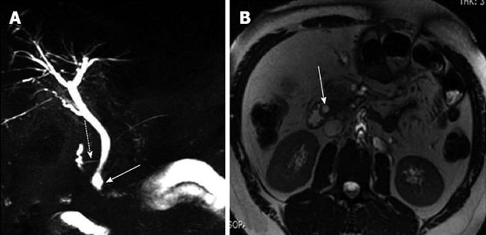
Figure 1 Choledochocele and associated pancreas divisum.
A: Magnetic resonance cholangiopancreatography showing the cystic dilatation of the terminal portion of the common bile duct (white arrow) in the setting of pancreas divisum (dashed arrow); B: Transverse view, white arrow indicates the common bile duct.
- Citation: Patidar Y, Agarwal N, Gupta S, Arora A, Mukund A, Rajesh S. Choledochocele with pancreas divisum: A rare co-occurrence diagnosed on magnetic resonance cholangiopancreatography. World J Radiol 2013; 5(7): 264-266
- URL: https://www.wjgnet.com/1949-8470/full/v5/i7/264.htm
- DOI: https://dx.doi.org/10.4329/wjr.v5.i7.264


