Copyright
©2013 Baishideng Publishing Group Co.
World J Radiol. May 28, 2013; 5(5): 226-228
Published online May 28, 2013. doi: 10.4329/wjr.v5.i5.226
Published online May 28, 2013. doi: 10.4329/wjr.v5.i5.226
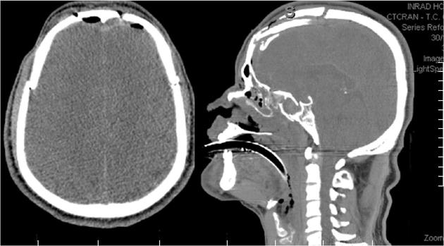
Figure 1 Computed tomography scan showed diffuse brain swelling with multiple skull fractures, including one in the anterior fossa.
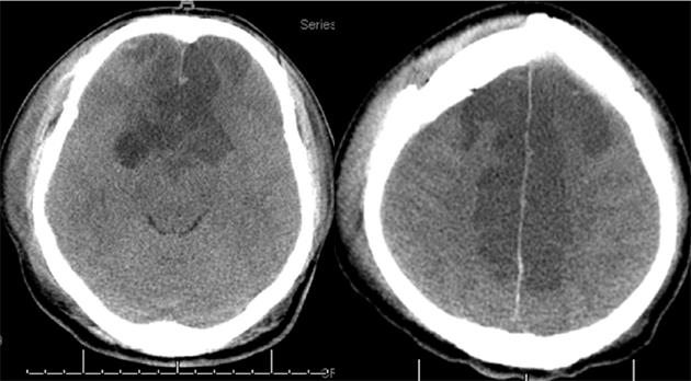
Figure 2 Follow up computed tomography scan showed extensive cerebral infarction in the territory of anterior cerebral artery.
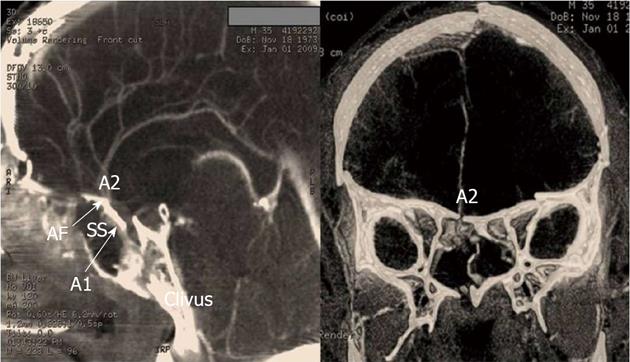
Figure 3 Multislice computed tomography angiography showed occlusion of the anterior cerebral artery adjacent to a fracture in the anterior fossa.
AF: Anterior Fossa; A1: First segment of anterior cerebral artery; A2: Second segment of anterior cerebral artery; SS: Sphenoid sinus.
- Citation: Paiva WS, de Andrade AF, Soares MS, Amorim RL, Figueiredo EG, Teixeira MJ. Occlusion of the anterior cerebral artery after head trauma. World J Radiol 2013; 5(5): 226-228
- URL: https://www.wjgnet.com/1949-8470/full/v5/i5/226.htm
- DOI: https://dx.doi.org/10.4329/wjr.v5.i5.226


