Copyright
©2013 Baishideng Publishing Group Co.
World J Radiol. Apr 28, 2013; 5(4): 184-186
Published online Apr 28, 2013. doi: 10.4329/wjr.v5.i4.184
Published online Apr 28, 2013. doi: 10.4329/wjr.v5.i4.184
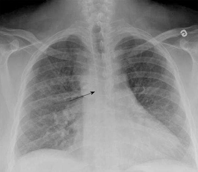
Figure 1 Chest X-ray scan showing the right aortic knob and arch as well as slight shift of the inferior trachea to the left (arrow).
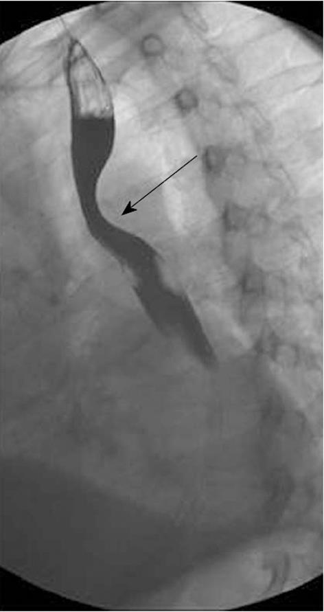
Figure 2 Barium esophagogram showing the posterior compression of the esophagus (arrow) in the oblique view.
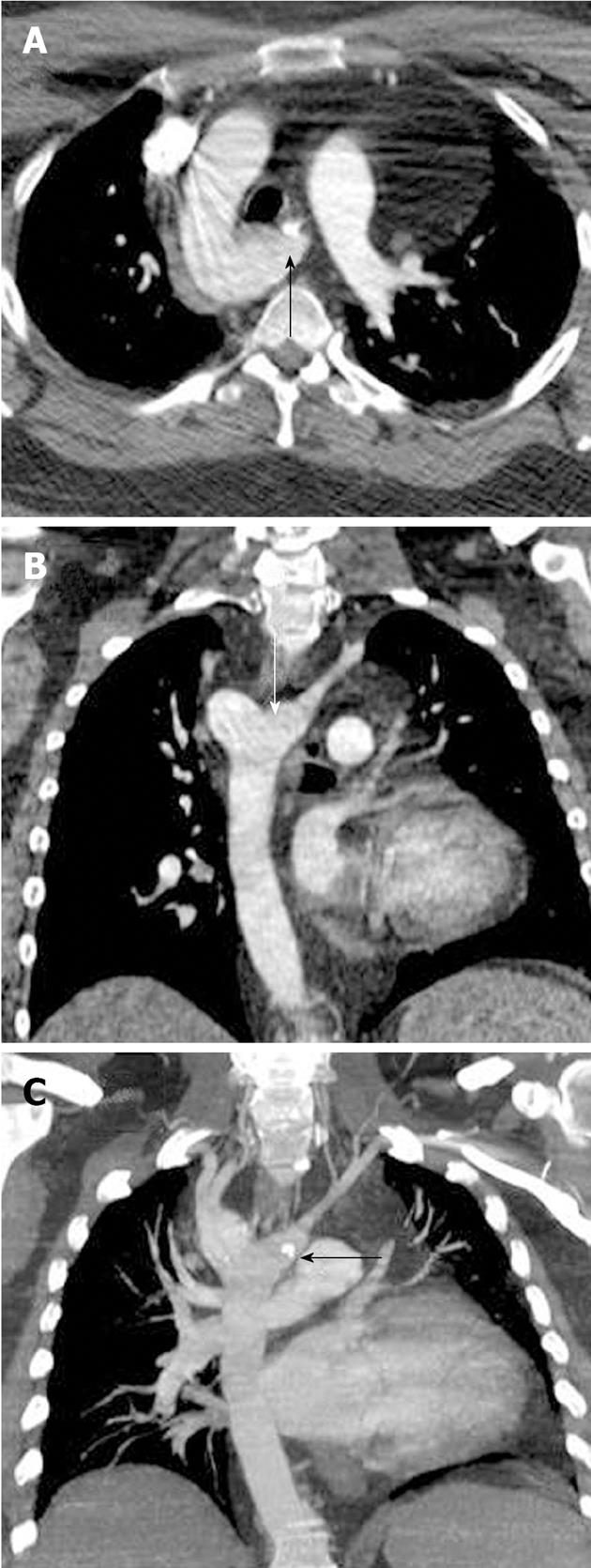
Figure 3 Multidetector computed tomography of the right aortic arch with an aberrant left subclavian artery in a 50-year-old man with dysphagia.
Axial (A) and coronal (B) multiplanar reformatted and maximum intensity projection (C) images showing the kommerell diverticulum (white arrow). Note the calcification in the presumed aortic insertion site of the ligamentum arteriosum (black arrows).
Figure 4 Non-contrast computed tomography in another patient.
A 30-year-old asymptomatic man. Axial (A), coronal (B) and sagittal (C), reformatted images demonstrating the course of the calcified ligament arteriosum (arrows).
- Citation: Kanza RE, Berube M, Michaud P. MDCT of right aortic arch with aberrant left subclavian artery associated with kommerell diverticulum and calcified ligamentum arteriosum. World J Radiol 2013; 5(4): 184-186
- URL: https://www.wjgnet.com/1949-8470/full/v5/i4/184.htm
- DOI: https://dx.doi.org/10.4329/wjr.v5.i4.184


