Copyright
©2013 Baishideng.
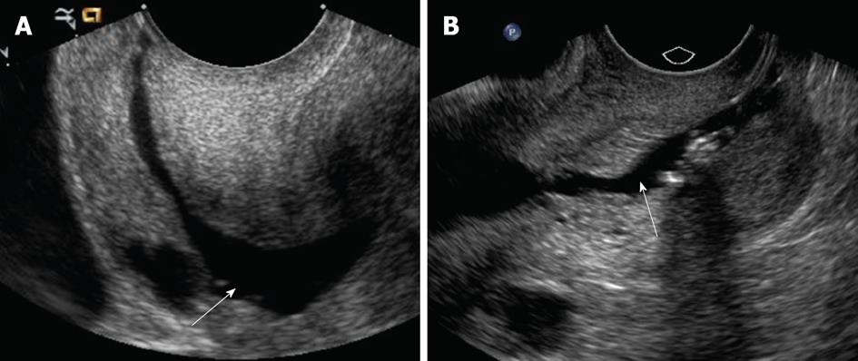
Figure 1 Procedural steps: Fluid instillation into the endometrial cavity (A, B).
Longitudinal ultrasound images of the lower uterine segment show adequate distention of the endocervical canal with warm saline during sonohysterography (arrows). During this phase, it is critical to observe for over or underdistention of the endocervical canal.
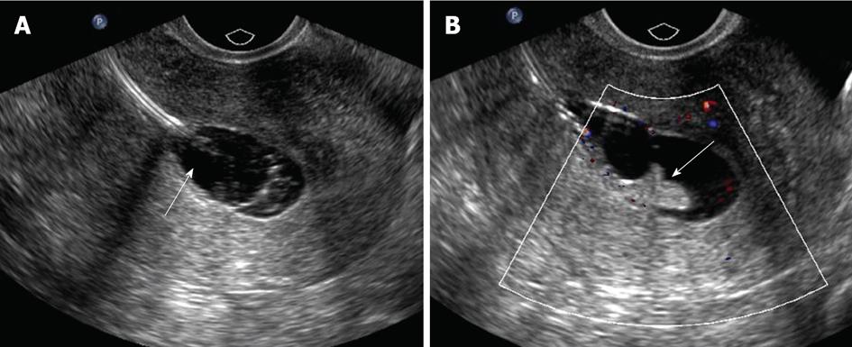
Figure 2 Common pitfalls during sonohysterography.
A: Longitudinal ultrasound image showing balloon hyperinflation during sonohysterography (arrow), which displaces and obscures an endometrial polyp; B: After the balloon is deflated to an adequate level, the echogenic endometrial polyp becomes evident (arrow).
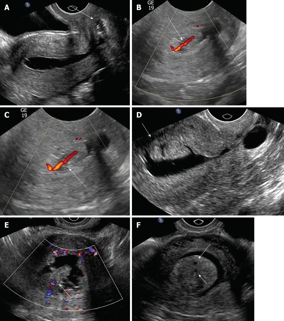
Figure 3 Multiple manifestations of endometrial polyps.
A: Sonohysterographical image of a cervical polyp (arrow); B, C: Sonohysterograms with Doppler demonstrating endometrial polyps with feeding vessels (arrows); D: Sonohysterogram shows elongated bilobed mass (arrow) attached to endometrial and projecting into the endometrial canal, representing an endometrial polyp; E: Broad based, hypoechoic, slightly hypervascular mass with slightly jagged borders projecting into the endometrial canal (arrow); F: Sonohysterogram shows single, echogenic polypoid lesion, representing an endometrial polyp arising with several tiny cysts (arrow).
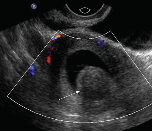
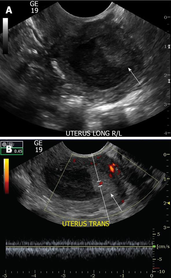
Figure 4 Doppler sonohysterogram showed a submucosal fibroid, manifested by broad based heterogenous, largely hypoechoic soft tissue projection (arrow) in the endometrial canal.
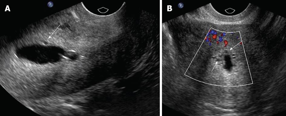
Figure 5 Endometrial hyperplasia as demonstrated by mild proliferation (arrows) of the endometrium on sonohysterogram (A, B).
This finding, however, is nonspecific.
Figure 6 Sonohysterogram which show masses (arrows) of heterogeneous echotexture within the endometrial cavity, consistent with endometrial cancer (A, B).
Figure 7 Sonohysterogram shows anechoic foci (arrows) in the subendometrial region, consistent with adenomyosis (A, B).
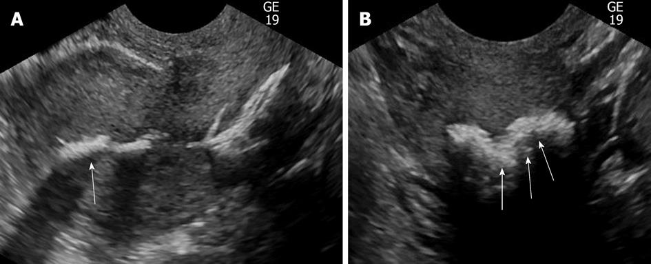
Figure 8 Sonohysterography demonstrating irregular hyperechoic linear densities (arrows) with acoustic shadowing involving the endometrium, consistent with osseous metaplasia (A, B).
- Citation: Yang T, Pandya A, Marcal L, Bude RO, Platt JF, Bedi DG, Elsayes KM. Sonohysterography: Principles, technique and role in diagnosis of endometrial pathology. World J Radiol 2013; 5(3): 81-87
- URL: https://www.wjgnet.com/1949-8470/full/v5/i3/81.htm
- DOI: https://dx.doi.org/10.4329/wjr.v5.i3.81


