Copyright
©2013 Baishideng.
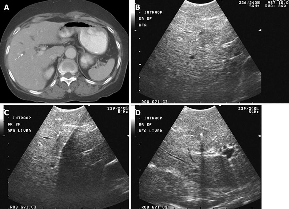
Figure 1 A 79-year-old male with metastatic colon with rising carcinoembryonic antigen and new hypodense lesion in the right liver on computed tomography examination.
A: Axial contrast-enhanced computed tomography shows a small hypodense lesion at the junction of segments VIII and V, concerning for metastasis; B: Intraoperative transverse sonogram identifies a solid slightly hypoechoic lesion in the right liver, at the junction of segments VIII-V, compatible with a metastasis; C, D: Longitudinal and transverse intraoperative ultrasound image shows the position of a radiofrequency ablation needle, with its in appropriate location within the center of the lesion. Intraoperative ultrasound is extremely useful to precise localize hepatic lesions and guide therapeutic interventions.
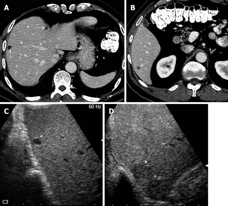
Figure 2 A 73-year-old male with colorectal cancer and indeterminate hepatic lesion on computed tomography, suspicious for metastasis.
A: Axial contrast-enhanced computed tomography (CT) image shows small hypodense lesions in the right liver, possibly representing cysts; B: Axial contrast-enhanced CT image more inferiorly in the same patient reveals a subcentimeter lesion, deemed too small to be accurately characterized. Metastasis was not excluded; C: Intraoperative ultrasound image in the longitudinal plane shows a homogeneously hypoechoic lesion in the dome of the liver consistent with a cyst; D: Longitudinal Intraoperative ultrasound image more inferiorly shows a solid hypoechoic lesion in the inferior right liver, consistent with a metastasis.
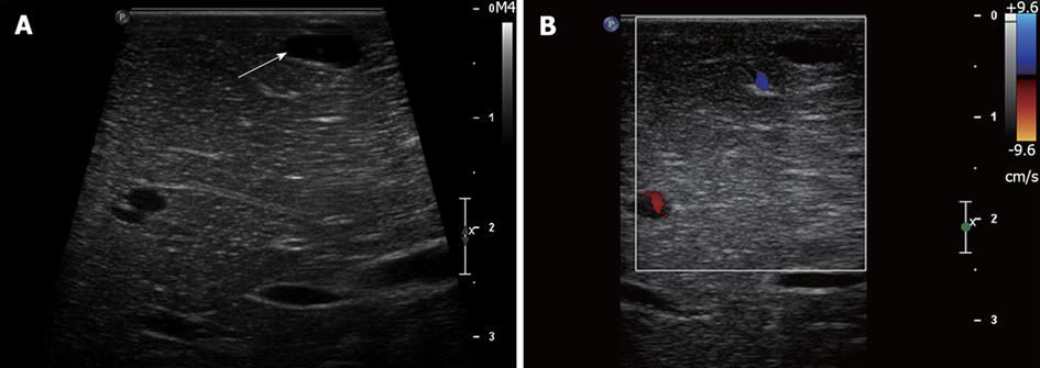
Figure 3 A 60-year-old male with potentially resectable pancreatic cancer and indeterminate liver lesions on preoperative imaging.
A: Intraoperative ultrasound image shows a homogeneously hypoechoic subcentimeter lesion in the left liver consistent with a cyst; B: Intraoperative ultrasound image with Doppler interrogation shows lack of internal vascularity, confirming the benign nature of this cyst.
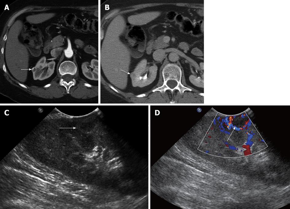
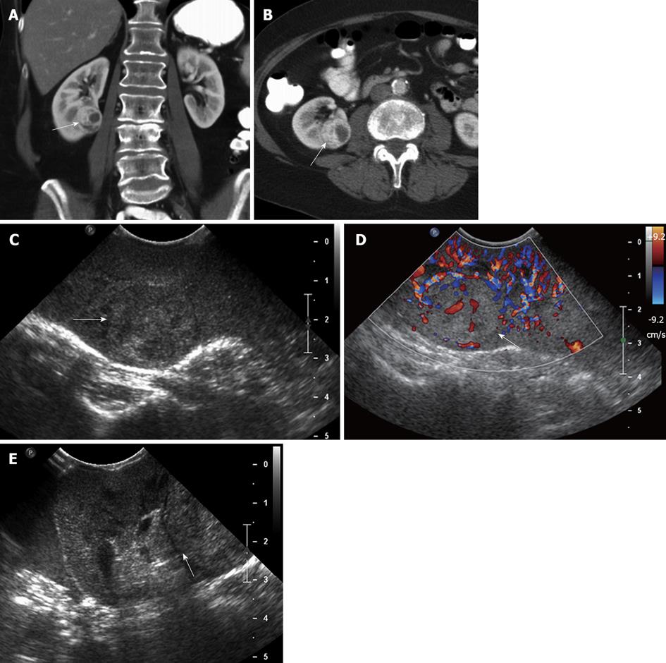
Figure 4 A 51-year-old female with incidentally detected small solid mass in the right kidney, compatible with a renal cell carcinoma.
A, B: Axial contrast-enhanced images of the right kidney show a 1.2 cm hypervascular mass in the midportion of the right kidney, concerning for a small renal cell carcinoma; C: Longitudinal Intraoperative ultrasound image localizes the small solid mass in the midportion of the right kidney anteriorly; D: Intraoperative ultrasound with Doppler interrogation shows prominent vessels at the margin of the lesion. Intraoperative ultrasound is an invaluable resource to localize small solid renal lesions during partial nephrectomy, ensuring that negative margins are achieved, while preserving the normal renal parenchyma.
Figure 5 A 62-year-old female with renal cell carcinoma.
A, B: Coronal (A) and axial (B) contrast-enhanced computed tomography images show a solid, hypervascular, 2.8 cm mass in the midportion - lower pole of the right kidney, consistent with a renal cell carcinoma; C, D: Intraoperative grayscale (C) and color Doppler ultrasound (D) identify a solid echogenic mass in the midportion - lower pole of the right kidney, with evidence of internal vascularity, consistent with a renal cell carcinoma; E: Intraoperative ultrasound image in a oblique transverse plane shows that the lesion abuts the hyperechoic renal sinus fat. This important information to guide the surgeon, in order that tumor-free margins are properly obtained at the time of resection.
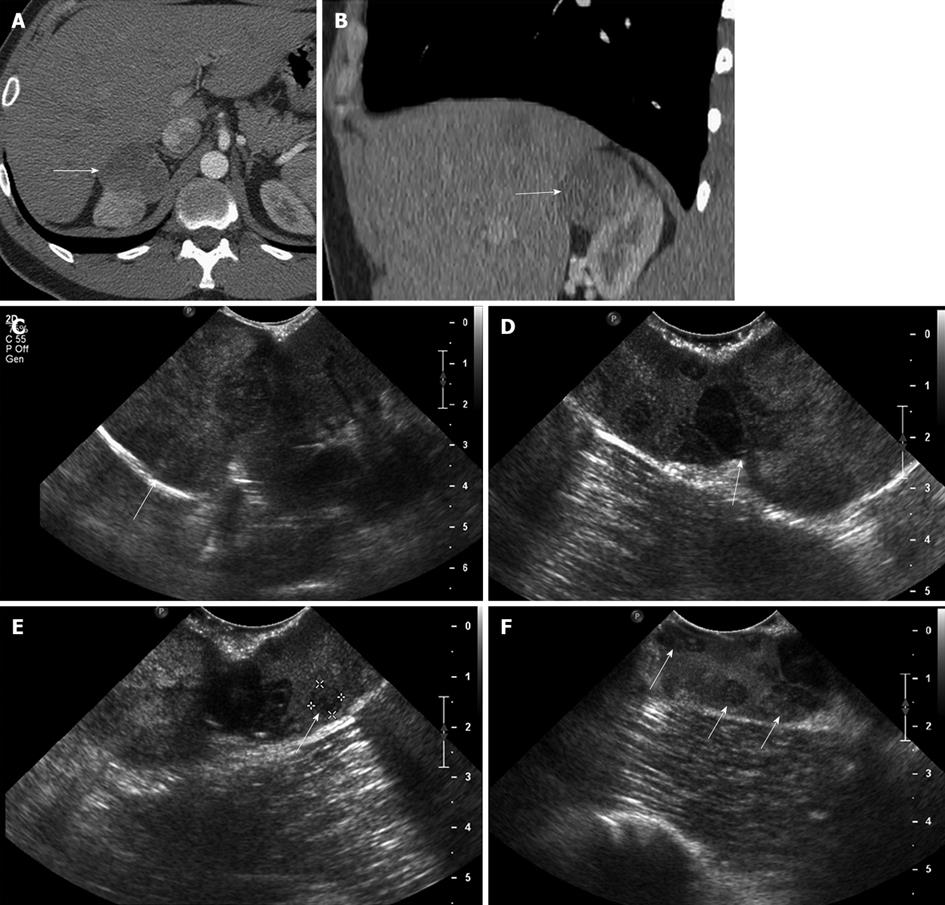
Figure 6 A 29-year-old male with renal cell carcinoma of the right kidney.
A, B: Axial (A) and coronal (B) contrast-enhanced computed tomography image show a large solid mass in the superior pole of the right kidney, consistent with a renal cell carcinoma; C: Intraoperative longitudinal sonogram shows an exophytic large solid mass in the superior pole of the right kidney; D: Intraoperative ultrasound image shows the point of attachment of the solid mass to the superior pole of the right kidney; E, F: High resolution intraoperative ultrasound image detects additional subcentimeter solid renal masses in the superior pole of the right kidney, near the site of origin of the large exophytic right renal mass. These findings alerted the surgeon that a deeper resection needed to be performed in order to secure tumor-free resection margin.
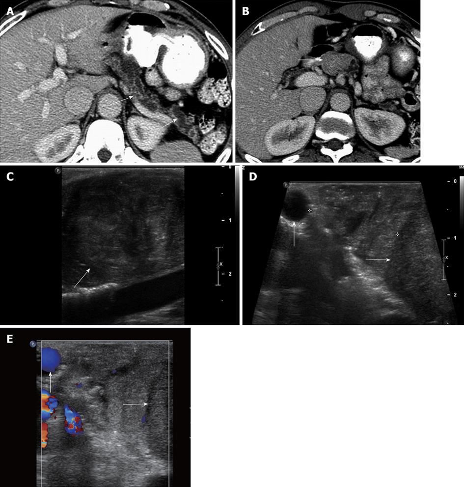
Figure 7 A 60-year-old male with pancreatic duct dilatation and suspected mass pancreatic head mass.
A: Axial contrast-enhanced computed tomography (CT) images show marked pancreatic ductal dilatation in the tail and body of the pancreas, with atrophy of the pancreatic parenchyma; B: Axial contrast-enhanced CT shows fullness in the region of the head of the pancreas, concerning for an isodense pancreatic head mass; C: Intraoperative ultrasound image clearly defines a solid mass in the head of the pancreas, consistent with a pancreatic ductal adenocarcinoma; D, E: Grayscale (D) and color Doppler (E) intraoperative ultrasound images show to better advantage the margins of the mass (arrow) in the cephalad head of the pancreas. The lesion is confined to the pancreas, and does not involve the gastroduodenal artery (vertical arrow). Intraoperative ultrasound is useful to assess size, margins of pancreatic mass, and their relationship with the adjacent vessels. This is particularly useful in cases of isodense pancreatic masses, which can be very difficult to evaluate with CT.
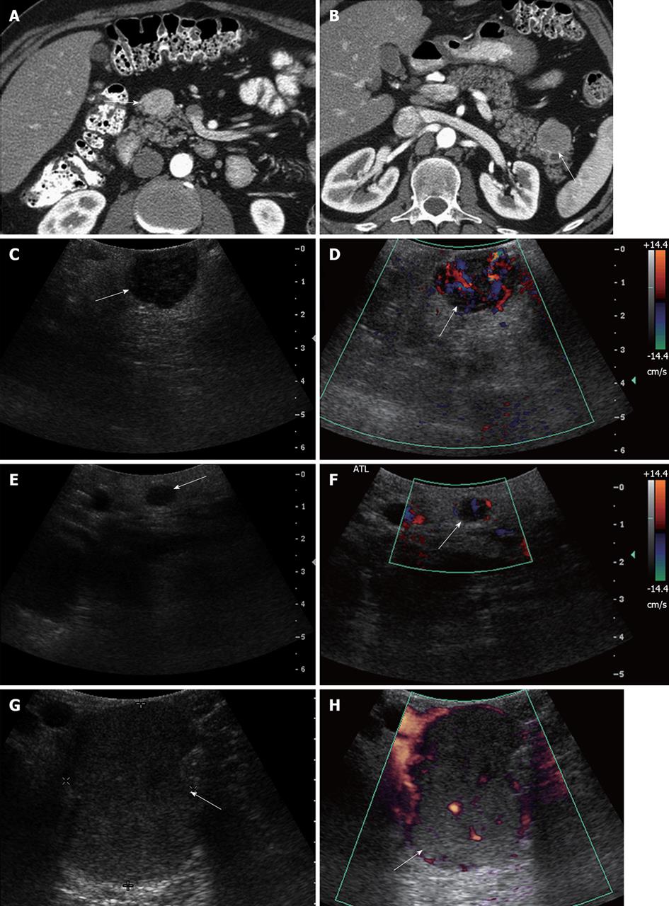
Figure 8 A 46-year-old male with multiple endocrine neoplasia type I syndrome and pancreatic neuroendocrine tumor.
A, B: Axial contrast-enhanced computed tomography (CT) images show hypervascular masses in the head and tail of the pancreas, consistent with neuroendocrine tumors; C: Intraoperative ultrasound image shows a well-defined solid mass in the head of the pancreas, consistent with the neuroendocrine tumor on CT; D: Doppler interrogation reveals increased vascularity within this mass; E, F: Intraoperative grayscale and color Doppler ultrasound images detect a 5 mm solid mass with internal vascularity in the head of the pancreas, consistent with a small neuroendocrine tumor. This lesion was not identified on the CT examination; G, H: Intraoperative grayscale and color Doppler ultrasound reveals the large dominant mass in the tail of the pancreas, consistent with a neuroendocrine tumor.
- Citation: Marcal LP, Patnana M, Bhosale P, Bedi DG. Intraoperative abdominal ultrasound in oncologic imaging. World J Radiol 2013; 5(3): 51-60
- URL: https://www.wjgnet.com/1949-8470/full/v5/i3/51.htm
- DOI: https://dx.doi.org/10.4329/wjr.v5.i3.51


