Copyright
©2013 Baishideng.
World J Radiol. Mar 28, 2013; 5(3): 106-112
Published online Mar 28, 2013. doi: 10.4329/wjr.v5.i3.106
Published online Mar 28, 2013. doi: 10.4329/wjr.v5.i3.106
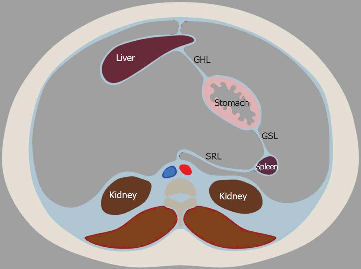
Figure 1 Embryologic development and rotation of the peritoneal structures to the eventual adult position.
The liver fills and moves to the right side of the peritoneal cavity as it grows. The spleen moves likewise to the left. GHL: Gastrohepatic ligament; GSL: Gastrosplenic ligament; SRL: Splenorenal ligament.
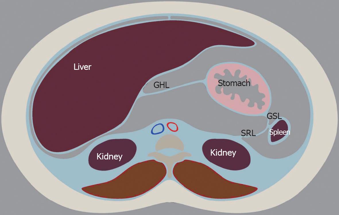
Figure 2 Peritoneal structures in the adult position after growth and rotation.
GHL: Gastrohepatic ligament; GSL: Gastrosplenic ligament; SRL: Splenorenal ligament.
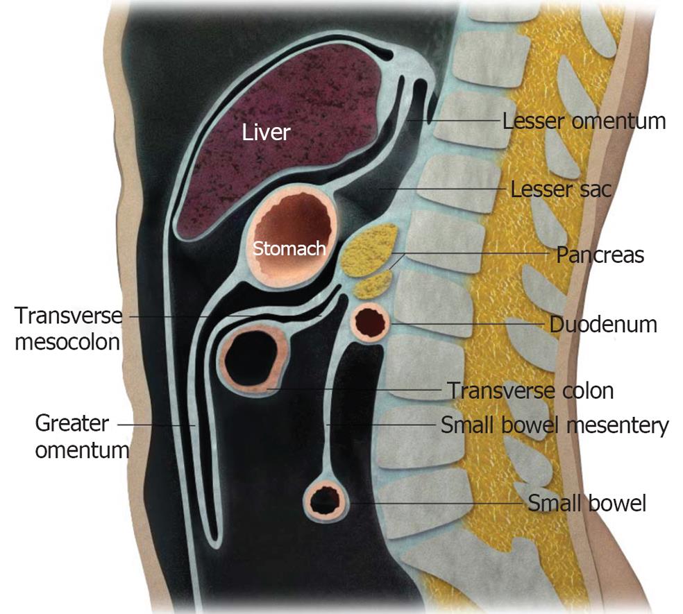
Figure 3 Diagrammatic representation of the peritoneal structures in sagittal view.
Figure 4 Coronal view of the peritoneal spaces and attachments within the supramesocolic and inframesocolic spaces.
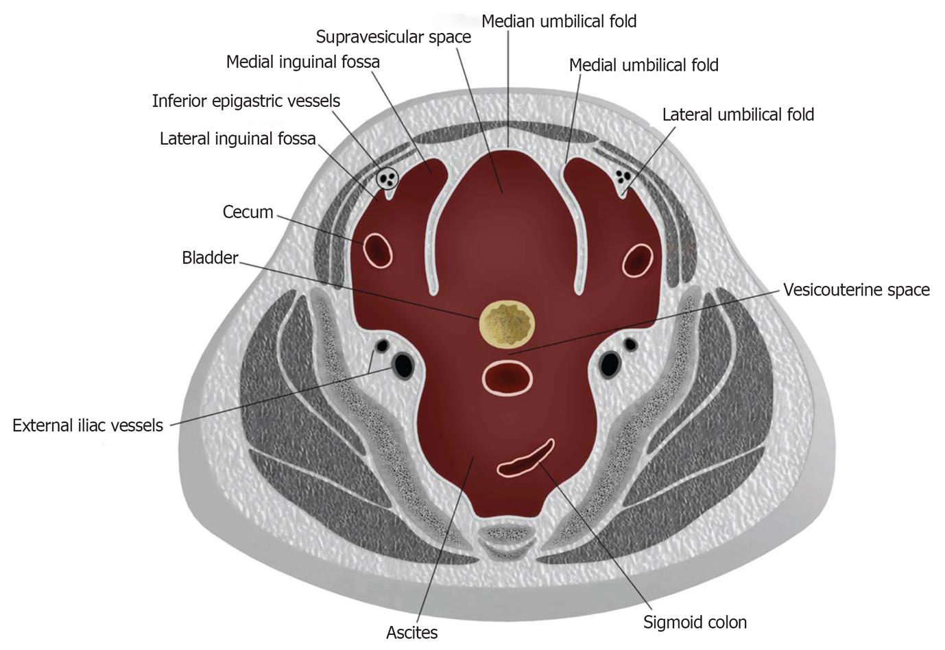
Figure 5 The paravesicular spaces in schematic cross section.
The anterior paravesicular space is divided into the supravesicular space, medial inguinal fossa and lateral inguinal fossa. The supravesicular space lies in between the medial umbilical fold. The lateral umbilical fold divides the medial and lateral inguinal fossa.
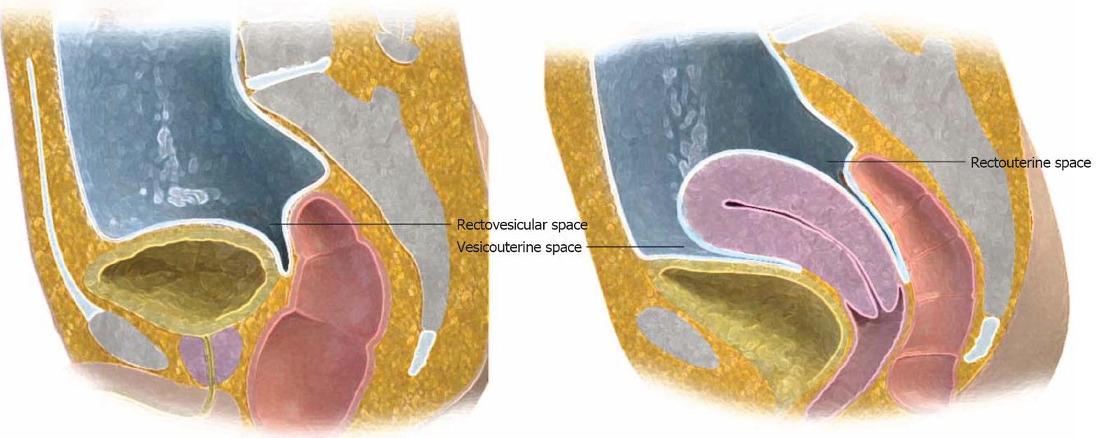
Figure 6 Posterior paravesicular spaces.
Sagittal view of the male (left) and female (right) pelvis demonstrates the peritoneal covering of the superior bladder, uterus and upper one-third of the rectum.
Figure 7 Computed tomography with intravenous contrast.
Thirteen years old with rhabdomyosarcoma. The anterior paravesicular spaces are well defined with ascites. The lateral umbilical fold (containing the inferior epigastric vessels, arrow) divides the lateral and medial inguinal fossa. In between the medial umbilical fold (containing the obliterated umbilical vein, arrowhead) is the supravesicular fossa.
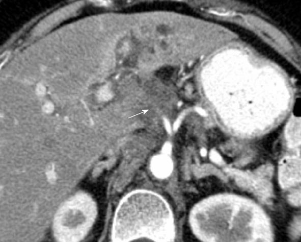
Figure 8 Computed tomography with intravenous contrast.
Metastatic gastric cancer involves a 66-year-old man. Tumor spreads along the gastrohepatic ligament from the stomach. This is evidenced by soft tissue infiltration of tumor along the left gastric artery, with loss of a normal fat plane (arrow).
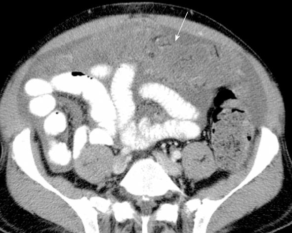
Figure 9 Computed tomography with intravenous contrast.
There is “omental caking” (arrow) in a patient with spread of colon cancer. Ascites is also present.
Figure 10 Computed tomography with intravenous contrast.
Intrahepatic cholangiocarcinoma spreads along the hepatoduodenal ligament in a 63-year-old female (arrow).
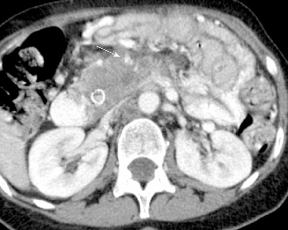
Figure 11 Computed tomography with intravenous contrast.
Metastatic disease spreads along the mesenteric root in a 62-year-old with pancreatic cancer. This is seen as soft tissue infiltration along the SMA (arrow). The SMV is occluded and not seen.
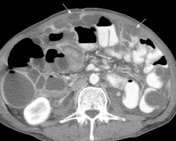
Figure 12 Computed tomography with intravenous contrast.
Pseudomyxoma peritonei from appendiceal carcinoma in a 66-year-old male seen as hypodense collections scalloping and exerting pressure on the surface of the bowel (arrows).
Figure 13 Schematic example of the ubiquitous lymphatic channels and nodes along the mesentery.
A mass in the rectosigmoid colon (labeled A) can spread superiorly along the lymphatic highway of the sigmoid mesocolon (arrow). Likewise, a mass located in the cecum can spread along the lymphatic channels of the ileocolic mesentery (labeled B).
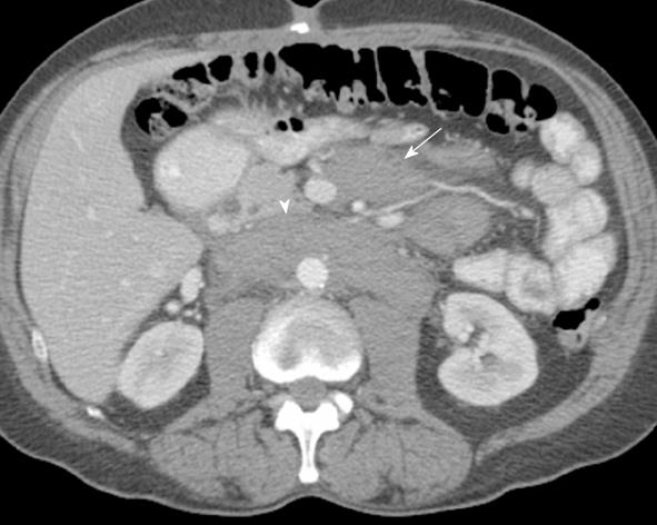
Figure 14 Computed tomography with intravenous contrast in a 66-year-old with lymphoma.
Enlarged lymph nodes along the mesentery represent lymphatic spread of disease (arrow). Retroperitoneal lymphadenopathy is also present (arrowhead).
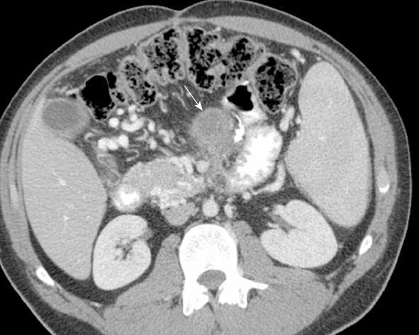
Figure 15 Computed tomography with intravenous contrast.
There is hematogenous spread of disease to the root of the mesentery in a 21-year-old with melanoma (arrow).
- Citation: Le O. Patterns of peritoneal spread of tumor in the abdomen and pelvis. World J Radiol 2013; 5(3): 106-112
- URL: https://www.wjgnet.com/1949-8470/full/v5/i3/106.htm
- DOI: https://dx.doi.org/10.4329/wjr.v5.i3.106


