Copyright
©2013 Baishideng Publishing Group Co.
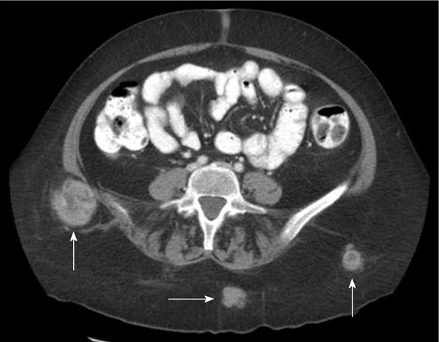
Figure 1 Subcutaneous implants.
Axial section from a contrast enhanced computed tomography demonstrates several enhancing masses (arrows) in the subcutaneous fat of this 62-year-old woman with metastatic melanoma.
Figure 2 Splenic metastasis.
A-C: Axial section from serial contrast enhanced computed tomography (CT) examinations over 1 year show an enlarging low attenuation lesion in the spleen (arrows). This was the only focus of metastatic disease in this 81-year-old woman with melanoma of the right ear. Note the smaller sattelite mass (dashed arrow, C) that has developed in the most recent CT.
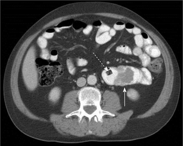
Figure 3 Small bowel metastasis.
Axial section from a contrast enhanced computed tomography in this 54-year-old man with metastatic melanoma shows a partially obstructing endolumenal mass (white arrow) in the jejunum. This patient presented with recurrent gastrointestinal bleeding. Note the video endoscopy capsule that failed to pass the metastatic implant (dashed arrow).
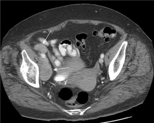
Figure 4 Pelvic metastasis.
Axial section from a contrast enhanced computed tomography in this 79-year-old woman with primary melanoma of the right heel shows enlarged right external iliac chain lymph nodes (arrows).
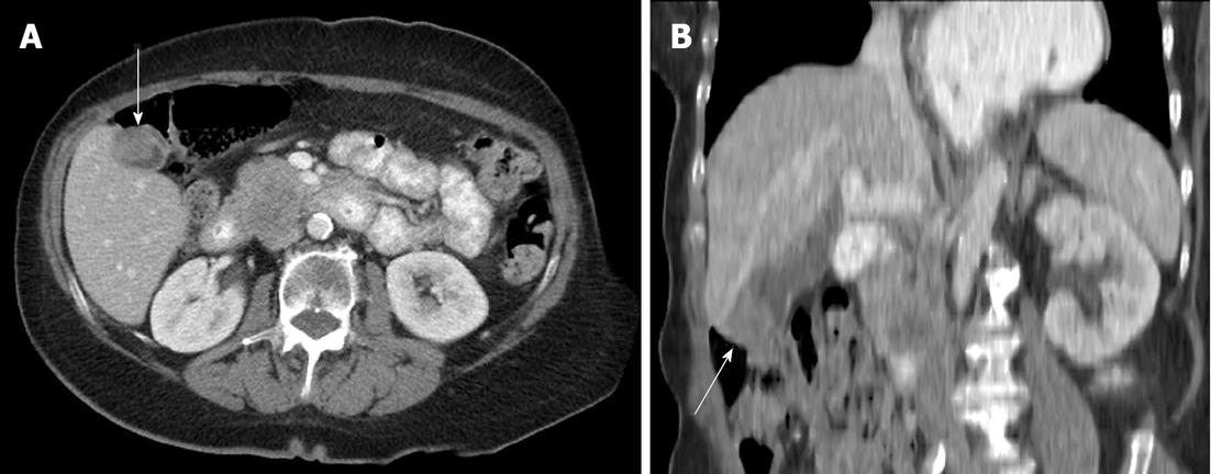
Figure 5 Gallbladder metastasis.
A, B: Axial section (A) and oblique reformat (B) from a contrast enhanced computed tomography in this 60-year-old woman with primary ocular melanoma shows a polypoid enhancing mass in the gallbladder fundus (arrows) consistent with a metastatic deposit. Metastases are also present in the pancreatic head and uncinate process.
Figure 6 Testicular metastasis.
A, B: Axial sections from a noncontrast computed tomography (CT) in this 41-year-old man with primary melanoma of the left flank show a mass at the superior pole of the right testis (arrow) consistent with a metastatic deposit. More normal appearing portions of the right testicle can be seen inferior to the mass (dashed arrow). Ultrasound confirmed the CT findings.
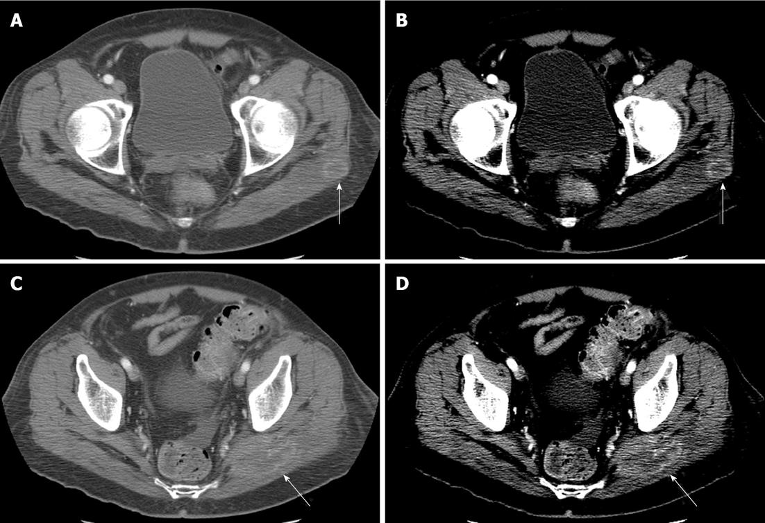
Figure 7 Skeletal muscle metastasis.
Axial sections from contrast enhanced computed tomography (CT) examinations in two different patients with metastases to the gluteus maximus (arrows). A, C: Images are displayed in typical soft tissue windows (ww/wl: 500/50); B, D: Images are the same acquired images displayed in more narrow windows (ww/wl: 200/50). Note the increased conspicuity of the lesions when narrow windows are used.
- Citation: Trout AT, Rabinowitz RS, Platt JF, Elsayes KM. Melanoma metastases in the abdomen and pelvis: Frequency and patterns of spread. World J Radiol 2013; 5(2): 25-32
- URL: https://www.wjgnet.com/1949-8470/full/v5/i2/25.htm
- DOI: https://dx.doi.org/10.4329/wjr.v5.i2.25


