Copyright
©2013 Baishideng Publishing Group Co.
World J Radiol. Oct 28, 2013; 5(10): 356-371
Published online Oct 28, 2013. doi: 10.4329/wjr.v5.i10.356
Published online Oct 28, 2013. doi: 10.4329/wjr.v5.i10.356
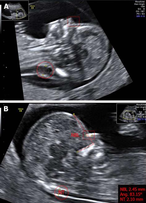
Figure 2 Markers of chromosomal defects.
A: Fetus with Down’s syndrome: increased NT (red circle), and absent nasal bone (red square where nasal bone was expected) at 11 wk of pregnancy; B: Normal fetus: measurements of nuchal translucency (NT, red circle), facial angle (red dashed line) and nasal bone length (NBL, red square) at 13 wk of pregnancy. The image has been certified by the Fetal Medicine Foundation. Photos taken by Wolfgang Moroder. Creative Commons[113].
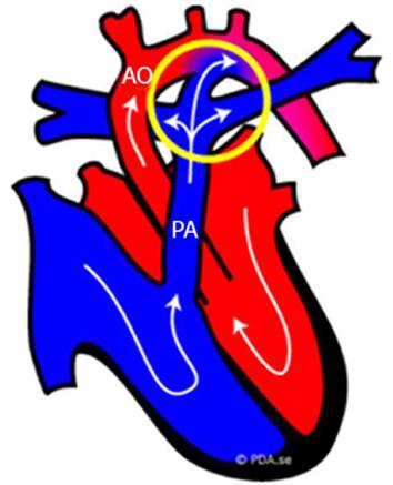
Figure 3 Fetal normal heart.
Schematic representation of the blood circulation in fetus: the blood that comes into the right side of the fetal heart (blue part) is pumped into the pulmonary trunk (PA) and flows through the ductus arteriosus (circled in yellow) directly out into the aorta (AO). The ductus arteriosus is an extra blood vessel of the fetal heart that creates a bypass for the blood oxygenated not by the lungs, but through the placenta[114].
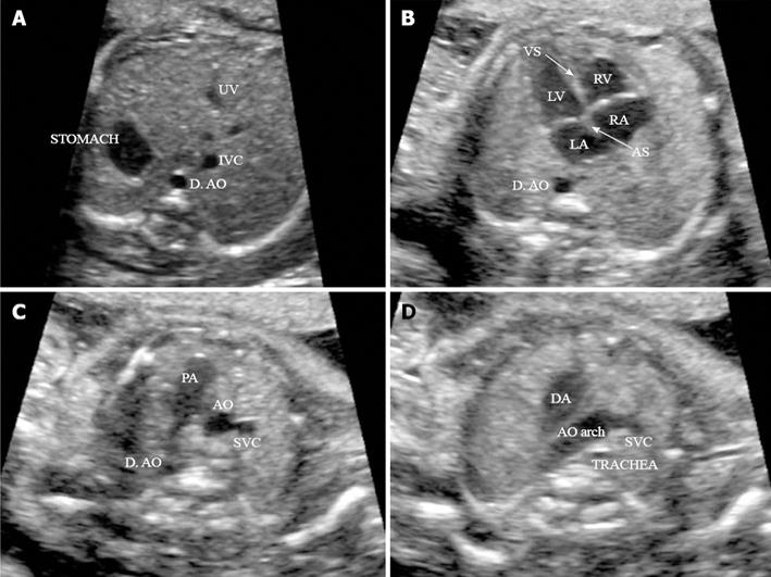
Figure 4 Four routine axial views of heart and great vessels.
A: Transverse view of the superior abdomen: the stomach on the fetal left side; the descending aorta (D. AO) to the left side and inferior vena cava (IVC) to right side of the spine, respectively; B: Four-chamber view: in a normal fetal heart, approximately equal size of the right and left chambers, intact intact ventricular septum (VS) and normal offset of the two atrioventricular valve; C: Three-vessel view: pulmonary artery (PA), aorta (AO) and superior vena cava (SVC) in the correct position and alignment; PA, to the left, is the largest of the three and the most anterior, whereas the SVC is the smallest and most posterior; D: Transverse view of the aortic arch: in the normal heart, both the AO arch and the ductal arch (DA) are located to the left of the trachea, in a ‘V’-shaped configuration. (Adapted from ISUOG Practice Guidelines[115]). UV: umbilical vein; RV: right ventricle; LV: left ventricle; LA: left atrium; RA: right atrium; AS: Atrial septum.
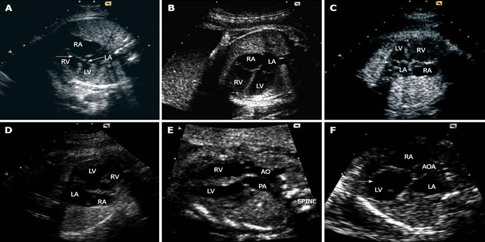
Figure 5 Markers of congenital heart disease.
A: Atrioventricular septal defect with a common junction leading to loss of off-setting of the atrioventricular valves (arrows); B: Four-chamber echo view in a normal mid-trimester fetus; C: Enlarged coronary sinus seen as a circular structure (arrow) within the left atrium adjacent to the mitral valve; D: Pulmonary atresia with intact ventricular septum: apical muscle bundles are prominent in the apical portion of the right ventricle with trabeculations coarser than usual; E: Transposition of the great arteries (discordant ventriculoarterial connections): the arteries are parallel to one another with the aorta arising from the right ventricle and positioned to the right of the pulmonary trunk; F: Severe aortic stenosis with patent mitral valve: the left ventricle becomes bulb-shaped (arrow). LA: left atrium; RA: right atrium; RV: right ventricle; LV: left ventricle; AO and AOA: Aorta; PA: Pulmonary trunk. Adapted from Cook et al[74].
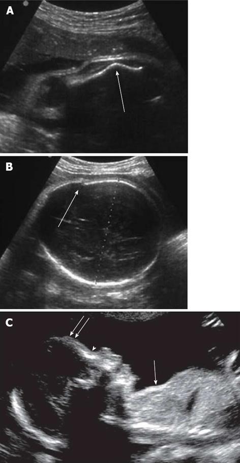
Figure 6 Marker of skeletal dysplasia by two-dimensional ultrasound.
A: Abnormal angulated femur; B: Osteogenesis imperfecta: cranial vault distortion upon probe pressure. (Adapted from Cassart[98]); C: Features of thanatophoric dysplasia: depressed nasal bridge (arrowhead), prominent forehead (double arrows), and undersized thorax (single arrow) compared with the abdomen. Adapted from Dighe et al[99].
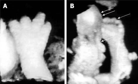
Figure 7 Skeletal anomalies detected by three-dimensional ultrasonography.
A: Trident configuration of the digits and brachydactyly suggestive of achondroplasia; B: Facial dysmorphisms: frontal bossing (double arrows) and flattened mid-face (single arrow), disproportionate limb segments and brachydactyly (dotted arrow) typical of achondroplasia. Adapted from Krakow et al[108].
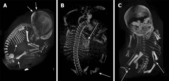
Figure 8 Prenatal diagnosis by three-dimensional helical computer tomography.
A: Achondroplasia (sagittal view): macrocephaly (double arrows), short ribs (dotted arrow) and increased thickness of the femoral metaphysis (single arrow); B: Osteogenesis imperfecta (posterior view): fractures of ribs and femur (arrows); C: Chondrodysplasia punctata (frontal view): epiphyseal calcifications of long bones (arrows). Adapted from Ruano et al[102].
- Citation: Renna MD, Pisani P, Conversano F, Perrone E, Casciaro E, Renzo GCD, Paola MD, Perrone A, Casciaro S. Sonographic markers for early diagnosis of fetal malformations. World J Radiol 2013; 5(10): 356-371
- URL: https://www.wjgnet.com/1949-8470/full/v5/i10/356.htm
- DOI: https://dx.doi.org/10.4329/wjr.v5.i10.356


