Copyright
©2012 Baishideng Publishing Group Co.
World J Radiol. Sep 28, 2012; 4(9): 413-417
Published online Sep 28, 2012. doi: 10.4329/wjr.v4.i9.413
Published online Sep 28, 2012. doi: 10.4329/wjr.v4.i9.413
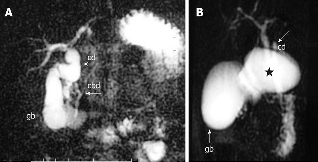
Figure 1 Magnetic resonance cholangiopancreatography.
A: Fusiform dilatation of the cystic duct; B: Saccular dilatation of the proximal cystic duct (star). Arrow points to normal caliber distal cystic duct. gb: Gallbladder; cd: Cystic duct; cbd: Common bile duct.
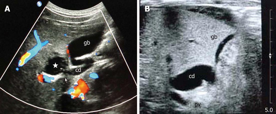
Figure 2 Ultrasonography.
A: Focal saccular dilatation of the cystic duct (star), arrow points to communication of the cyst with cystic duct; B: Mild fusiform dilatation of the cystic duct. gb: Gallbladder; cd: Cystic duct.
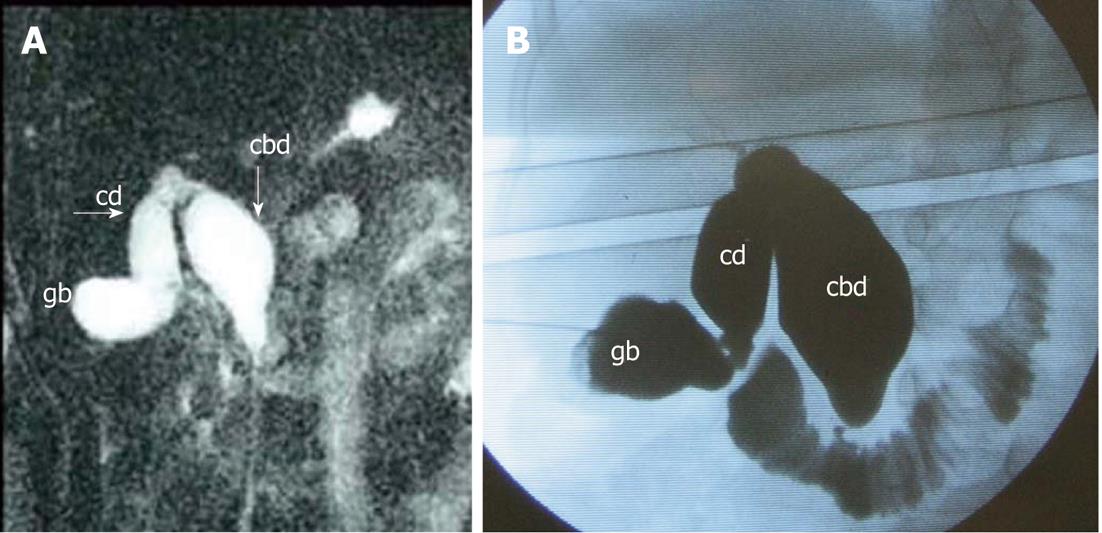
Figure 3 Magnetic resonance cholangiopancreatography (A) and intraoperative cholangiography (B) shows fusiform dilatation of the common bile duct and cystic duct.
gb: Gallbladder; cd: Cystic duct; cbd: Common bile duct.
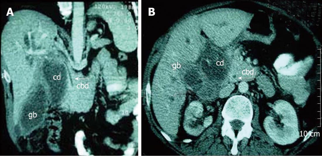
Figure 4 Coronal multiplanar reformatted (A) and axial (B) computed tomography scan shows saccular dilatation of the cystic duct and abnormal soft tissue suggesting malignancy.
White arrow points to normal common bile duct. Black arrow points to abnormal soft tissue suggesting malignancy within dilated cystic duct. gb: Gallbladder; cd: Cystic duct; cbd: Common bile duct.
- Citation: Maheshwari P. Cystic malformation of cystic duct: 10 cases and review of literature. World J Radiol 2012; 4(9): 413-417
- URL: https://www.wjgnet.com/1949-8470/full/v4/i9/413.htm
- DOI: https://dx.doi.org/10.4329/wjr.v4.i9.413


