Copyright
©2012 Baishideng Publishing Group Co.
World J Radiol. Jul 28, 2012; 4(7): 341-344
Published online Jul 28, 2012. doi: 10.4329/wjr.v4.i7.341
Published online Jul 28, 2012. doi: 10.4329/wjr.v4.i7.341
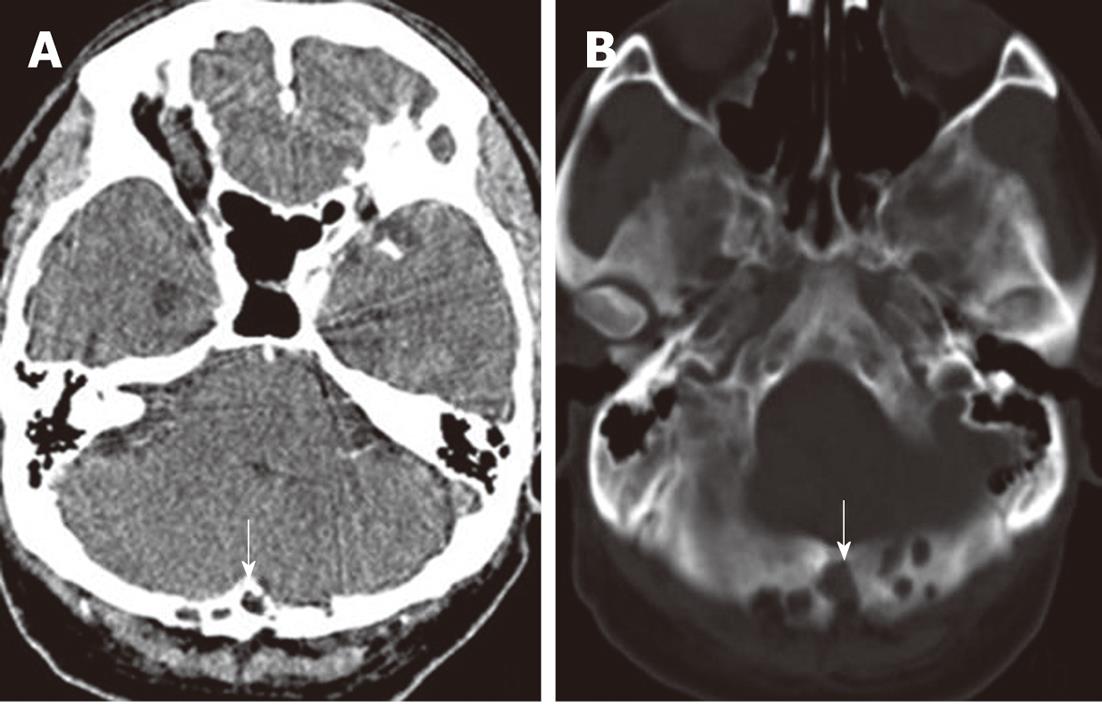
Figure 1 Multiple arachnoid granulations (arrows) in a 56-year-old man.
Axial noncontrast computed tomography on soft-tissue windows (A) and bone windows (B) showed occipital multiple osteolytic lesions adjacent to the transverse sinus.
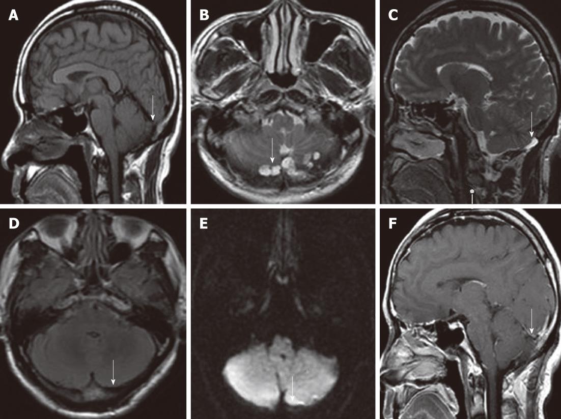
Figure 2 Multiple arachnoid granulations (arrows) in a 56-year-old man.
A-C: Sagittal T1-weighted image (T1WI) (A) showed hypointensity relative to the brain, axial (B) and sagittal (C) T2-weighted image showed hyperintensity relative to the brain, with a banded lower signal most likely representing the intra-arachnoid granulations (AGs) collagenous connective tissue; D: Axial fluid attenuated inversion recovery image demonstrated complete suppression of intra-AG fluid; E, F: Axial diffusion-weighted imaging (E) showed no restricted diffusion. Sagittal postcontrast T1WI (F) showed no gadolinium enhancement.
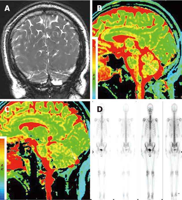
Figure 3 Multiple arachnoid granulations (arrow) in a 56-year-old man.
A: Coronal three-dimensional fast imaging employing steady state acquisition showed that the subarachnoid cavity communicated with a large arachnoid granulation (AG); B, C: Sagittal T2-mapping demonstrated T2 relaxation times of intra-AG contents paralleled that of cerebrospinal fluid in the ventricles and adjacent subarachnoid spaces; D: Single photon emission computed tomography with 99mTc-Methyl diphosphonate revealed no increased radiotracer accumulation.
- Citation: Lu CX, Du Y, Xu XX, Li Y, Yang HF, Deng SQ, Xiao DM, Li B, Tian YH. Multiple occipital defects caused by arachnoid granulations: Emphasis on T2 mapping. World J Radiol 2012; 4(7): 341-344
- URL: https://www.wjgnet.com/1949-8470/full/v4/i7/341.htm
- DOI: https://dx.doi.org/10.4329/wjr.v4.i7.341


