Copyright
©2012 Baishideng Publishing Group Co.
World J Radiol. Jul 28, 2012; 4(7): 324-327
Published online Jul 28, 2012. doi: 10.4329/wjr.v4.i7.324
Published online Jul 28, 2012. doi: 10.4329/wjr.v4.i7.324
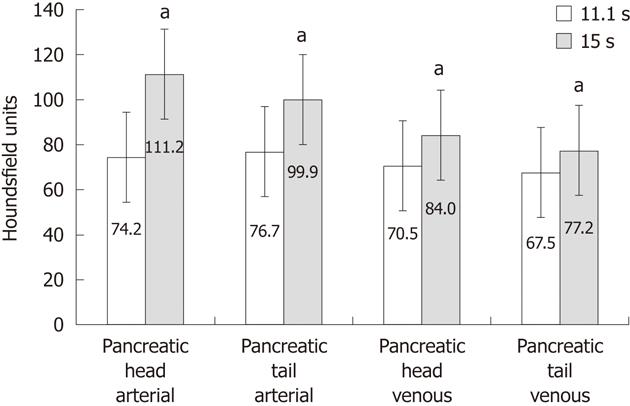
Figure 1 Influence of multidetector computed tomography scan delay on mean attenuation levels of pancreatic parenchyma.
aP < 0.05.
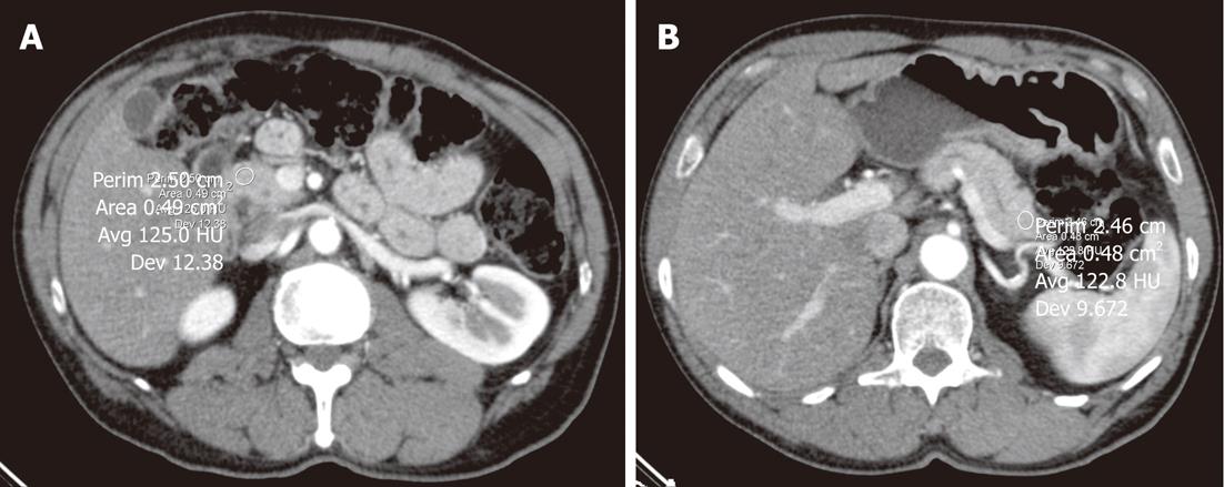
Figure 2 Axial arterial phase multidetector computed tomography image in a 58-year-old male patient with weight loss.
A:The scan delay for multidetector computed tomography examination was 15 s. The mean attenuation level was 125.0 HU in the pancreatic head; B: Within the parenchyma of the pancreatic tail, the mean attenuation value was 122.8 HU.
- Citation: Stuber T, Brambs HJ, Freund W, Juchems MS. Sixty-four MDCT achieves higher contrast in pancreas with optimization of scan time delay. World J Radiol 2012; 4(7): 324-327
- URL: https://www.wjgnet.com/1949-8470/full/v4/i7/324.htm
- DOI: https://dx.doi.org/10.4329/wjr.v4.i7.324


