Copyright
©2012 Baishideng Publishing Group Co.
World J Radiol. May 28, 2012; 4(5): 193-207
Published online May 28, 2012. doi: 10.4329/wjr.v4.i5.193
Published online May 28, 2012. doi: 10.4329/wjr.v4.i5.193
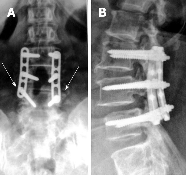
Figure 1 Antero-posterior (A) and lateral digital radiographs (B) of the lumbosacral spines with posterior lumbar interbody fusion showing LV3 through LV5 levels laminectomies with fine posteriorly located bone chips (arrows) on antero-posterior view that are difficult to appreciate on lateral view.
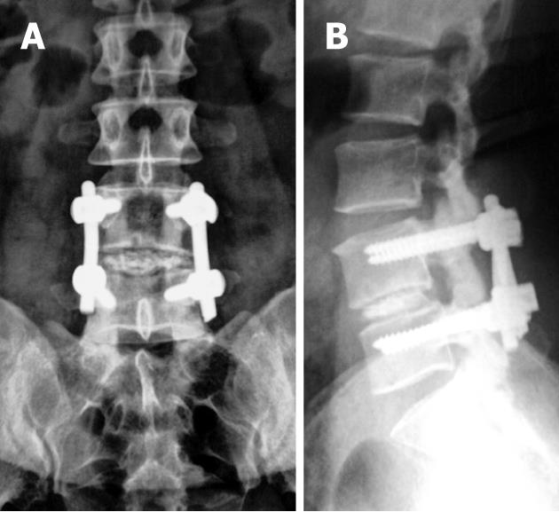
Figure 2 Immediate post-operative antero-posterior (A) and lateral digital radiographs (B) of the lumbosacral spines with posterior lumbar interbody fusion showing LV4-LV5 levels laminectomies with iliac bone graft at LV4-5 disc level.
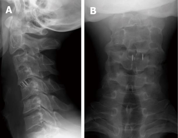
Figure 3 Antero-posterior (A) and lateral radiographs (B) of the cervical spines showing metallic markers of CV4-5 intervertebral disc spacers.
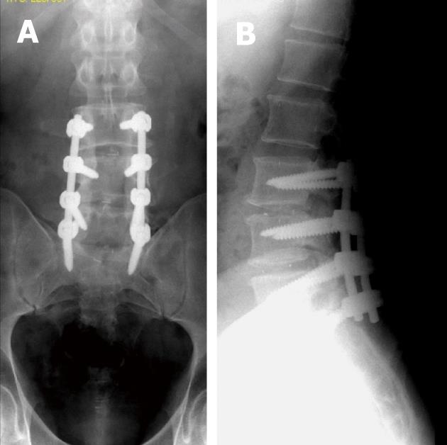
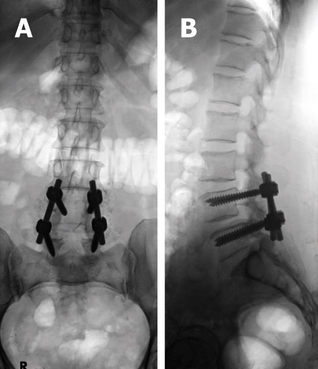
Figure 4 Antero-posterior (A) and lateral digital radiographs (B) of the lumbosacral spines with posterior lumbar interbody fusion showing LV3 through SV1 levels laminectomies with 2 metallic marks denoting insertion of radiolucent intervertebral disc spacers.
Note the posterior mark is at least 2 mm anterior to posterior border of adjacent vertebral body at LV3-4 level.
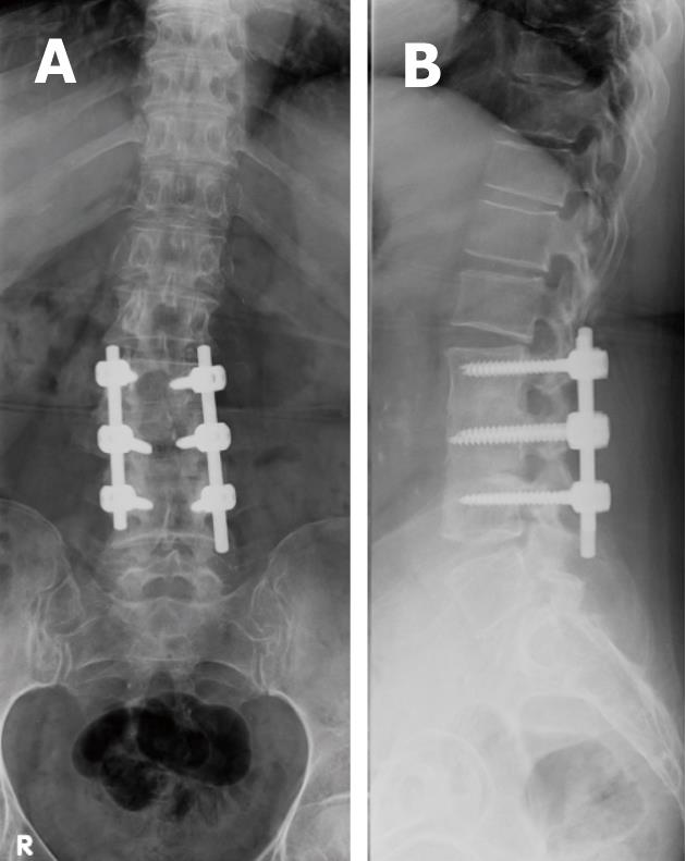
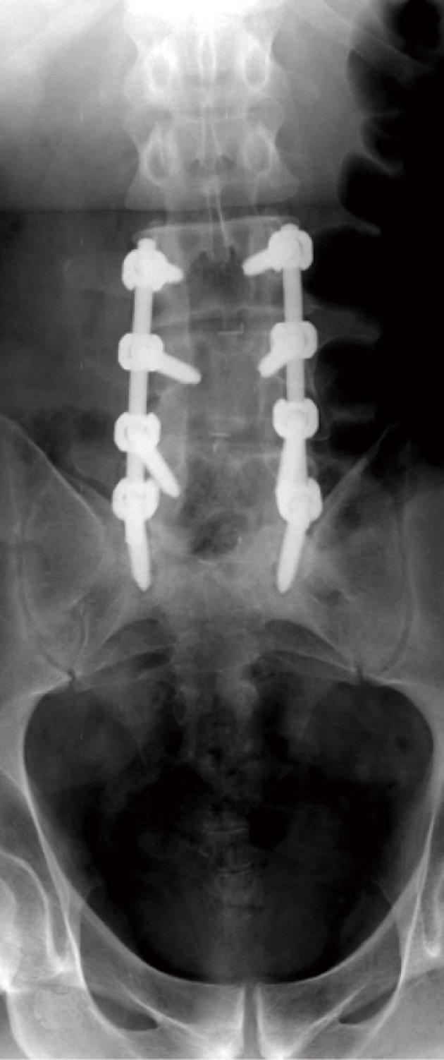
Figure 5 Antero-posterior (A) and lateral radiographs (B) showing well positioned spinal lumbar construct with pedicular screws central within the pedicles and not violating the cortices or adjacent endplates.
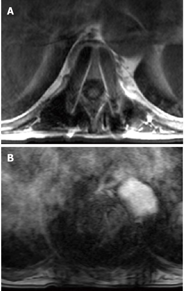
Figure 6 T1W SE image (A) demonstrating magnetic resonance susceptibility artifact of metallic hardware are mainly the sum of signal loss within the metallic object and high signal intensity appearing around the metallic object caused by altered both phase and frequency of local spins leading to read-out misregistration.
This is more obtrusive on T2-gradient images (B).
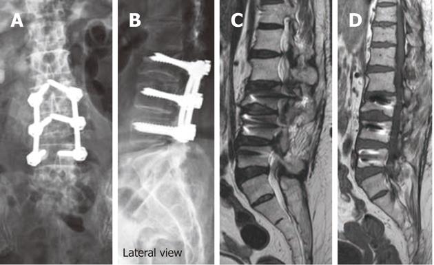
Figure 7 Antero-posterior (A) and lateral digital radiographs (B) of the lumbosacral spines showing aberrant pedicular screw violating LV3 superior endplate and protruding into LV2-3 disc, T2W (C) and T1W (D) sagittal magnetic resonance images showing aberrant pedicular screw violating LV3 superior endplate and protruding into LV2-3 disc.
Figure 8 Fluoroscopic spot views of the lumbo-sacral spines depicting the fine bone chips used for fusion beside the short-segment hardware at LV4 and LV5 levels.
These were barley seen on radiographic films.
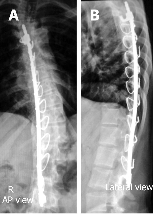
Figure 9 Antero-posterior (A) and lateral radiographs (B) of the dorso-lumbar spines showing a Harrington rod spanning the upper dorsal and lumbar vertebra for correction of adolescent scoliosis.
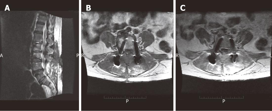
Figure 10 Sagittal T2W SE image (A), axial T1 pre-contrast (B) and post-contrast SE MR images (C) in a post-operative patient showing localized encysted collection within the operative bed with appreciable contrast enhancement of the encysted collection following IV gadolinium administration representing a post-operative infection with abscess formation.
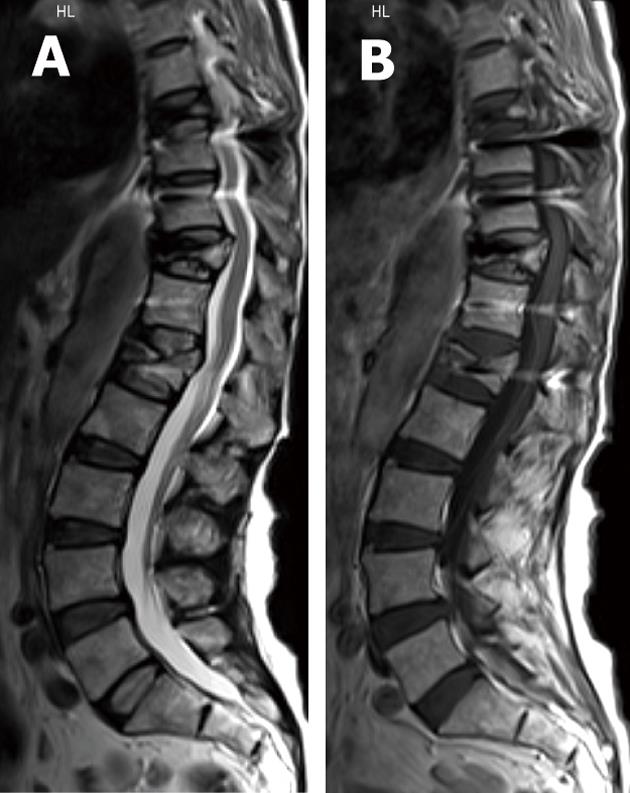
Figure 11 T2W SE (A) and T1W (B) sagittal magnetic resonance images showing multiple vertebral body collapse targeting LV1, DV11 and DV8 levels in osteoporotic patient predisposing to hardware failure.
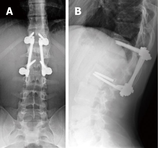
Figure 12 Antero-posterior (A) and lateral digital radiographs (B) of the lumbosacral spines with short-segment posterior fixation spanning LV5 and SV1 levels with fracture of left lower pedicular screw of the construct representing hardware failure.
Figure 13 Antero-posterior digital radiographs of the lumbosacral spines with posterior lumbar interbody fusion showing LV3 through SV1 levels laminectomies with peri-screw lucenecy seen around the last screws representing aseptic loosening.
- Citation: Nouh MR. Spinal fusion-hardware construct: Basic concepts and imaging review. World J Radiol 2012; 4(5): 193-207
- URL: https://www.wjgnet.com/1949-8470/full/v4/i5/193.htm
- DOI: https://dx.doi.org/10.4329/wjr.v4.i5.193


