Copyright
©2012 Baishideng Publishing Group Co.
World J Radiol. Apr 28, 2012; 4(4): 167-173
Published online Apr 28, 2012. doi: 10.4329/wjr.v4.i4.167
Published online Apr 28, 2012. doi: 10.4329/wjr.v4.i4.167
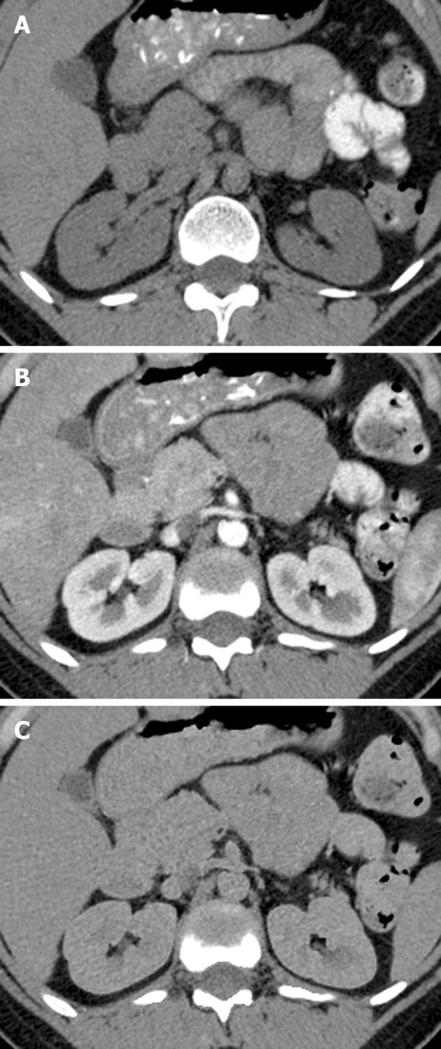
Figure 1 A 52-year-old female with the kidney contrast deletion.
A, B: The regular nonenhanced (A) and contrast-enhanced (B) images demonstrate kidneys with normal appearance; C: Virtual nonenhanced shows substantial deletion of contrast from the kidney parenchyma.
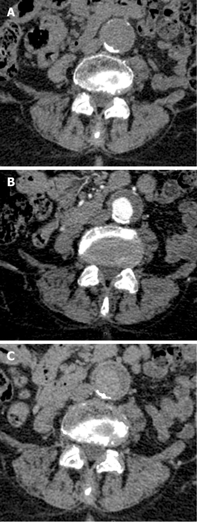
Figure 2 A 60-year-old male with an abdominal aortic aneurysm.
A: Regular nonenhanced image demonstrates a curved calcification in the posterior border of the aneurysm with some smaller calcifications in the anterior border; B: The contrast enhanced image shows opacification of the lumen; C: Virtual nonenhanced shows preserved posterior calcifications with deletion of the smaller anterior calcifications.
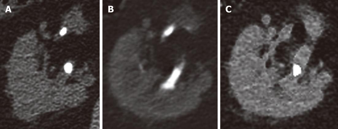
Figure 3 Regular nonenhanced image (A) demonstrates two stones in the right kidney of a 64-year-old male.
B: In the contrast-enhanced image, contrast excretion into the collecting system is inhibited by the stones, which are obscured; C: In the virtual nonenhanced image, the posterior stone is maintained while the anterior stone is deleted.
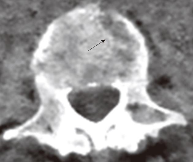
Figure 4 Virtual nonenhanced image of this 58-year-old female shows a lytic-like artifact in the vertebral body.
The “lesion” was easily identified as an artifact.
- Citation: Sosna J, Mahgerefteh S, Goshen L, Kafri G, Aviram G, Blachar A. Virtual nonenhanced abdominal dual-energy MDCT: Analysis of image characteristics. World J Radiol 2012; 4(4): 167-173
- URL: https://www.wjgnet.com/1949-8470/full/v4/i4/167.htm
- DOI: https://dx.doi.org/10.4329/wjr.v4.i4.167


