Copyright
©2012 Baishideng Publishing Group Co.
World J Radiol. Apr 28, 2012; 4(4): 151-158
Published online Apr 28, 2012. doi: 10.4329/wjr.v4.i4.151
Published online Apr 28, 2012. doi: 10.4329/wjr.v4.i4.151
Figure 1 A 31-year-old female with liver malignant fibrous histiocytoma.
A: Plain computed tomography scan shows well demarcated mass with direct contact to the left lobe of liver and uniform density of 38 HU; B: Contrast scan arterial phase shows heterogeneous enhancement of mass compare to normal liver parenchyma with surrounding blood vessels; C: Pathological specimen HE (× 400) showing multinucleated spindle shape tumor cells with inflammatory cells.
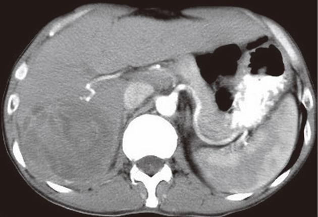
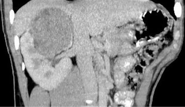
Figure 2 A 47-year-old male with liver malignant fibrous histiocytoma.
Contrast-enhanced computed tomography scan arterial phase shows a mass with a clear boundary in the right-posterior lobe of liver segment 6 and 7. No obvious enhancement with areas of hypodensity is evident.
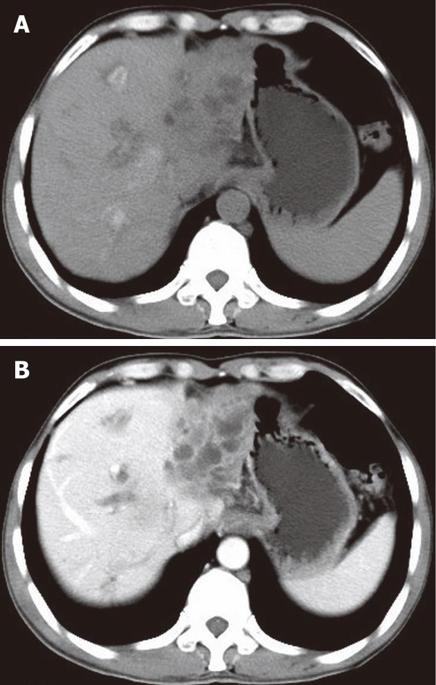
Figure 3 A 54-year-old male with liver malignant fibrous histiocytoma.
A: Non-contrast computed tomography shows ill defined lesion of density 30 HU in the left lobe with dilated intrahepatic biliary duct; B: Contrast scan portal vein phase shows low enhancement compared to normal liver parenchyma and multiple areas of low attenuation.
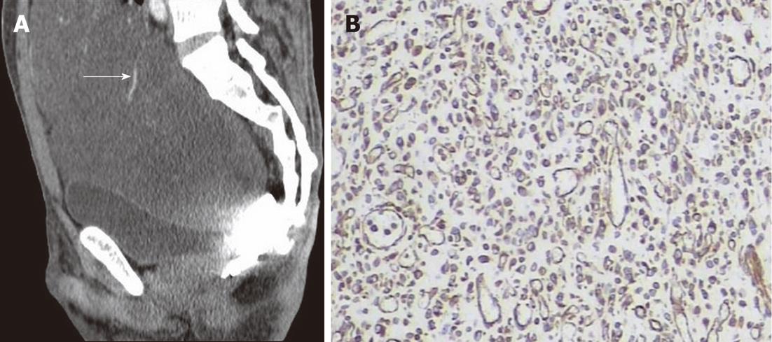
Figure 4 A 34-year-old male with greater omentum malignant fibrous histiocytoma.
A: Contrast-enhanced computed tomography scan sagittal view shows a well demarcated cystic mass extending from the abdominal to the pelvic cavity, note the blood vessel on the surface (arrow); B: Immunohistochemistry shows positive staining to vimentin (× 400).
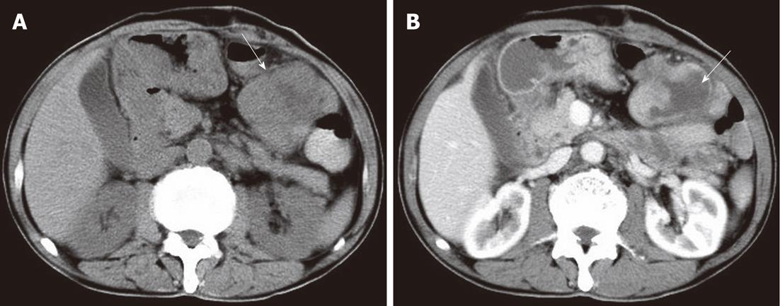
Figure 5 A 64-year-old male with superior mesentery malignant fibrous histiocytoma.
A: Non-contrast computed tomography scan shows mass (arrow) in the peritoneal cavity of 33 HU with a clear margin and central area of hypodensity; B: Contrast-enhanced scan venous phase shows mild enhancement with more pronounced central necrotic zone (arrow).
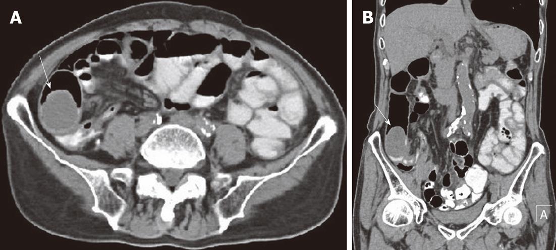
Figure 6 A 79-year-old male with ileum malignant fibrous histiocytoma, plain computed tomography scan abdomen.
A, B: Axial (A) and coronal view (B) show intraluminal and intramural soft tissue mass of density 34 HU in the ilio-cecal region (arrows) without significant areas of necrosis. Note the oral contrast stagnation in the proximal loop of the small intestine and dilatation of the ascending colon.
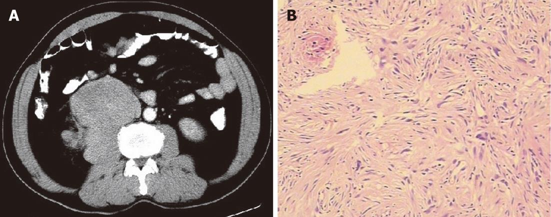
Figure 7 A 59-year-old male with retroperitoneal psoas muscle malignant fibrous histiocytoma.
A: Contrast-enhanced computed tomography scan arterial phase shows mass in retroperitoneum with clear margin, no obvious enhancement of mass and area of low density corresponding to necrosis. Note that the fat plane between the mass and psoas muscle is not evident, and the mass seems to be in direct contact with the right psoas muscle; B: Pathological specimen HE stain (× 400) shows spindle to oval tumor cells arranged in a criss-cross fashion.
Figure 8 A 23-year-old male with right kidney malignant fibrous histiocytoma.
Non-contrast computed tomography scan coronal view shows soft tissue mass of 47 HU in the upper pole of the right kidney extending toward the renal pelvis.
- Citation: Karki B, Xu YK, Wu YK, Zhang WW. Primary malignant fibrous histiocytoma of the abdominal cavity: CT findings and pathological correlation. World J Radiol 2012; 4(4): 151-158
- URL: https://www.wjgnet.com/1949-8470/full/v4/i4/151.htm
- DOI: https://dx.doi.org/10.4329/wjr.v4.i4.151


