Copyright
©2012 Baishideng Publishing Group Co.
World J Radiol. Apr 28, 2012; 4(4): 135-140
Published online Apr 28, 2012. doi: 10.4329/wjr.v4.i4.135
Published online Apr 28, 2012. doi: 10.4329/wjr.v4.i4.135
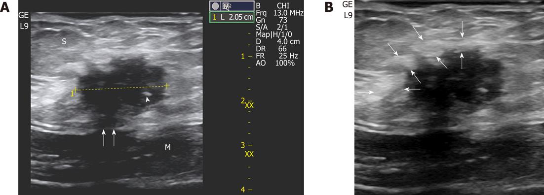
Figure 1 A 32-year-old-woman with 6-mo of cyclic pain two years after a cesarean delivery.
A: A small (2 cm) scar endometrioma is displayed at sonography located in the subcutaneous fat (S) with typical features: a roundish nodule, hypoechoic with fibrotic spots (the thick one is indicated by a white arrowhead), spiculated margins infiltrating the sheath (white arrows) of the rectus abdominis (M); B: An inflammatory hyperechoic ring (between white arrows) circumscribes almost the entire endometrioma.
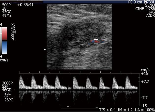
Figure 2 A small endometrioma with the typical single vascular pedicle entering the mass at the periphery.
No central vascularisation is observed. Doppler demonstrates high resistance arterial flow (resistive index > 0.7).
Figure 3 A 30-year-old woman with a long history (84 mo) of continuous pain in the lower abdomen and two previous non-diagnostic laparoscopic examinations.
In the abdominal wall (between the subcutaneous fat and the muscle), US exam discloses a 4-cm, ovoid hypoechoic endometrioma with a linear fistulous tract (arrow) emerging from the posterior aspect of the lesion and transgressing the muscular plane.
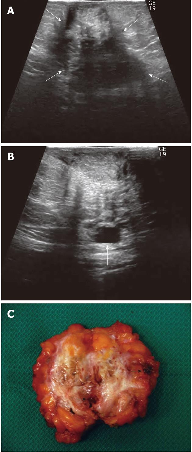
Figure 4 A 35-year-old woman with 52-mo of cyclic pain (after a cesarean delivery 2 years previously) that was not relieved by a surgical intervention for “intestinal adhesions” performed 44 mo before admission.
A: US displays a huge (widest diameter: 60 mm), heterogeneous, irregularly-shaped mass occupying (between white arrows) the entire abdominal subcutaneous fat thickness and infiltrating the underlying muscle; B: A cystic area is seen along the posterior aspect of the mass (arrow); C: Cut surface of the surgical specimen: note the irregular shape of the highly vascularised mass with margins infiltrating adjacent tissues, the huge amount of white fibrotic strands and the multiple, well-defined haemorrhagic cystic collections (black arrows point out the greatest collections).
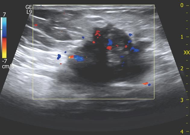
Figure 5 A 32-year-old woman with large scar endometrioma.
Color Doppler exam displays multiple vascular poles entering the mass from different points and intralesional vascularisation.
- Citation: Francica G. Reliable clinical and sonographic findings in the diagnosis of abdominal wall endometriosis near cesarean section scar. World J Radiol 2012; 4(4): 135-140
- URL: https://www.wjgnet.com/1949-8470/full/v4/i4/135.htm
- DOI: https://dx.doi.org/10.4329/wjr.v4.i4.135


