Copyright
©2012 Baishideng Publishing Group Co.
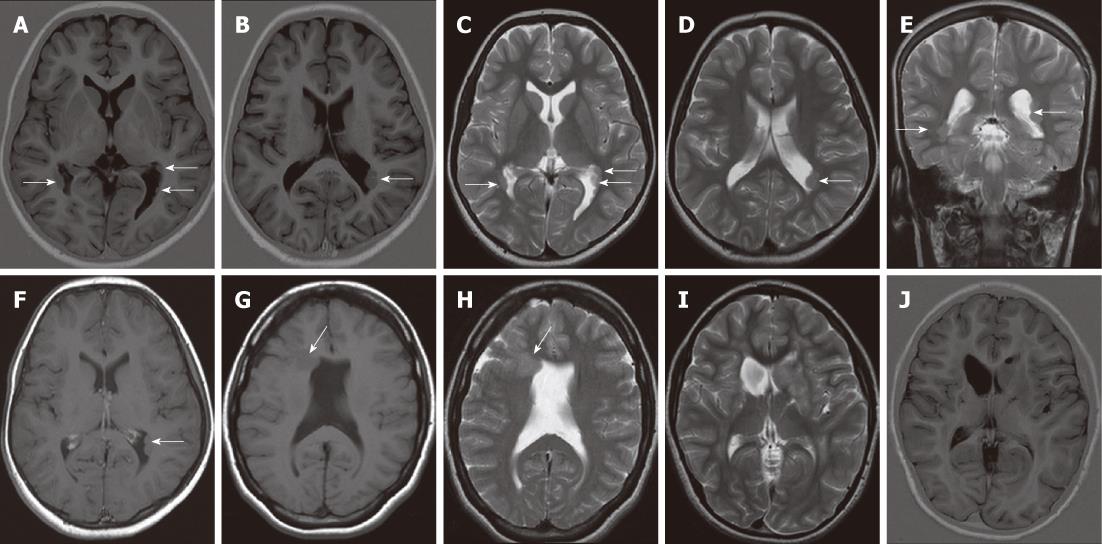
Figure 1 Subependymal heterotopia.
A, B: Axial inversion-recovery magnetic resonance (MR) images; C-E: Axial (C, D) and coronal T2 W (E) images showing multiple bilateral subependymal gray matter heterotopic nodules protruding into and indenting the trigone of the lateral ventricles and the left occipital horn (arrows). The nodules are isointense to cortical gray matter; F: Axial contrast-enhanced T1WI shows no enhancement of the nodules; G, H: Axial T1W and T2W MR images revealing unilateral focal subependymal gray matter nodule (arrow) protruding into and indenting the frontal horn of the right lateral ventricle. The nodule is isointense to the cortical gray matter. Mild ventricular dilatation is noted with absent septum pelucidum; I, J: Axial T2 WI and inversion-recovery MR image showing a unilateral, large subependymal heterotopic gray matter mass projecting into the frontal horn of the left lateral ventricle. The mass is isointense to the cortical gray matter.
Figure 2 Subcortical nodular heterotopia.
A: Contrast-enhanced axial computed tomography shows a large right fronto-parietal, non-enhancing mass exerting mass effect on the right lateral ventricle; B-D:Non-contrast enhanced magnetic resonance imaging (B), axial T1W, (C) axial T2W and (D) coronal T2W images showing a large subcortical nodular mass, isointense to the cortical gray matter. The overlying cortex is thin and the corpus callosum is agenetic.
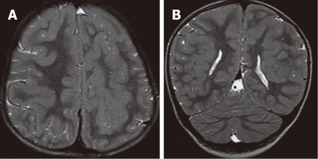
Figure 3 Subcortical curvilinear heterotopia.
A, B: Axial T2W and coronal T2W images showing bilateral curvilinear heterotopia within the white matter. The heterotopic tissue is convoluted and contiguous with the overlying cortex. Linear and punctuate cerebrospinal fluid signal are seen within the heterotopic tissue. The cerebral cortex shows pachygyria.
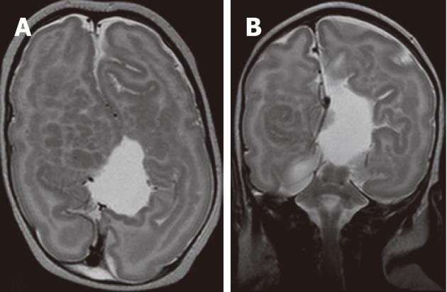
Figure 4 Mixed nodular and curvilinear subcortical heterotopia.
A, B: Axial T2W and coronal T2W images showing multiple nodular and curvilinear heterotopia within the white matter bilaterally. The overlying cortex shows pachygyria. The right cerebral hemisphere is smaller compared to the left cerebral hemisphere. The corpus callosum is agenetic with distorted lateral ventricles.
Figure 5 Band heterotopia.
A, B: Axial T2W and coronal inversion-recovery images showing bilateral thick bands of heterotopia, isointense to cortical gray matter within the white matter. Note the pachygyria and small left cerebral hemisphere; C, D: Axial T2W and inversion-recovery images showing bilateral, symmetric, continuous, smooth, thick bands of gray matter (arrow) outlined by thin layers of white matter and seems like a “double cortex”. Note the very thin, smooth cerebral cortex with absent cortical sulci (lissencephaly).
- Citation: Donkol RH, Moghazy KM, Abolenin A. Assessment of gray matter heterotopia by magnetic resonance imaging. World J Radiol 2012; 4(3): 90-96
- URL: https://www.wjgnet.com/1949-8470/full/v4/i3/90.htm
- DOI: https://dx.doi.org/10.4329/wjr.v4.i3.90


