Copyright
©2012 Baishideng Publishing Group Co.
Figure 1 Preliminary diagnostic imaging in Galen aneurysmal malformations patients.
A: Portable chest radiograph in a newborn (Patient 1) with clinical signs of congestive heart failure and a cranial bruit demonstrates cardiomegaly and pulmonary vascular congestion; B: A non-contrast head computed tomography shows an enlarged midline vascular structure in the posterior fossa consistent with a Galen aneurysmal malformations (VGAM); C, D: In another patient (Patient 3) who presented similarly, color Doppler ultrasound (C) indicates the presence of a posterior midline vascular pouch subsequently shown by magnetic resonance (MR) venography to be a VGAM (D); E, F: Alternatively, contrast-enhanced brain MR imaging was the initial diagnostic imaging used to diagnose a VGAM in Patient 2 who presented with macrocephalus and cranial bruit.
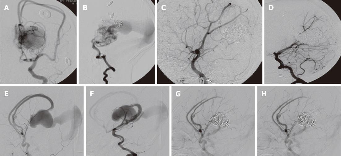
Figure 2 Catheter cerebral arteriography and embolization of Galen aneurysmal malformations.
A, B: Arteriovenous shunting through a fine arterial network is seen within choroidal-type Galen aneurysmal malformations (VGAMs); C, D: Combined transarterial and transvenous embolization in this patient (Patient 1) enabled complete obliteration of the lesion; E, F: Direct fistulas to the venous pouch with feeders from the anterior (E) and posterior (F) cerebral arteries are seen in mural-type VGAMs (E and F); G, H: Palliative partial embolization in this patient (Patient 3) enabled resolution of congestive heart failure.
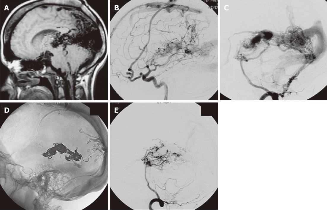
Figure 3 Galen aneurysmal malformations-associated cerebral vascular changes.
A: Sagittal T1-weighted magnetic resonance imaging in this adult (Patient 5) with a VGAM shows extensive flow voids within the quadrigeminal cistern and in the medial parietal and occipital lobes; B, C: Catheter cerebral arteriography with internal carotid (B) and vertebral artery (C) injections show that multiple independent arteriovenous fistulas of the sagittal sinus, falx, and tentorium are present; D, E: Post-embolization lateral scout (D) and left vertebral artery angiogram (E) show the coil mass within the venous pouch and near complete obliteration of the lesion.
- Citation: Ellis JA, Orr L, II PCM, Anderson RC, Feldstein NA, Meyers PM. Cognitive and functional status after vein of Galen aneurysmal malformation endovascular occlusion. World J Radiol 2012; 4(3): 83-89
- URL: https://www.wjgnet.com/1949-8470/full/v4/i3/83.htm
- DOI: https://dx.doi.org/10.4329/wjr.v4.i3.83


