Copyright
©2012 Baishideng Publishing Group Co.
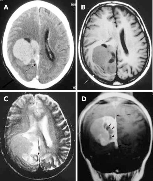
Figure 1 Solitary fibrous tumor.
Post-contrast computed tomography. A: A dural-based lobulated mass with dural tail enhancement (arrow); B: Axial T1-weighted magnetic resonance (MR) image shows an extra-axial mass, isointense to the gray matter compressing the adjacent cortical convolutions (arrow); C: Axial T2-weighted MR image shows that the mass has intermediate signal intensity. Note the peritumoral cysts with high signal intensity (arrow); D: On the post-contrast, T1-weighted MR image, the mass shows intense enhancement, intra-tumoral cysts (arrowheads) and dural tail sign (arrow).
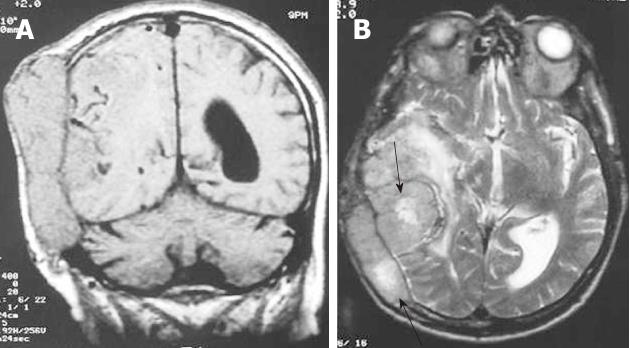
Figure 2 Hemangiopericytoma.
A: The coronal T1-weighted magnetic resonance (MR) image shows an extra-axial isointense mass with internal flow voids and calvarial destruction; B: On the axial, T2-weighted MR image the mass remains isointense with internal areas of hyperintensity and moderate peritumoral edema.
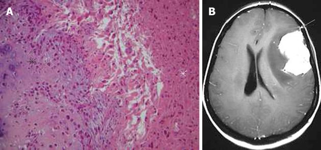
Figure 3 Gliosarcoma.
A: Pathological image showing an area with features of osteogenic sarcoma (black asterisk) adjacent to the glial component of the tumor (white asterisk) (hematoxylin-eosin, original magnification × 100); B: On the axial, post-contrast T1-weighted magnetic resonance image, the tumor shows intense homogeneous enhancement. Note the close relation of the mass with the dura, which is enhanced in a way mimicking meningioma (arrow).
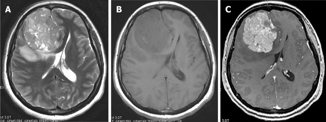
Figure 4 Dural metastasis in a 36-year-old woman with breast carcinoma.
A: Axial T2-weighted image shows a large extra-axial frontal heterogeneous mass; B: On T1-weighted magnetic resonance (MR) image the mass is isointense with the gray matter; C: Axial post-contrast T1-weighted MR image shows an extra-axial strongly enhanced lesion broad based to the falx mimicking meningioma.
Figure 5 Non-Hodgkin Lymphoma.
The axial non-contrast computed tomography. A: A hyperdense extra-axial mass extending to both sides of the frontoparietal bone; B: Bone window setting depict lytic lesion of the calvarium (arrow); C: On axial T2-weighted magnetic resonance images, the mass appears isointense with peritumoral edema; D: After the administration of contrast media, a strong homogeneous enhancement is seen with a dural tail (arrows).
Figure 6 Plasmocytoma in a 62-year-old patient.
A: Typical plasma cell morphology hematoxylin-eosin stain; B: Axial T2-weighted magnetic resonance images show an isointense frontal mass with peritumoral edema compressing the right ventricle; C: The axial post-contrast image shows intense homogeneous enhancement of the lesion with a dural tail (arrow).
Figure 7 Rosai-Dorfman disease.
A: The non-contrast computed tomography (CT) shows a peripheral, poorly marginated, hyperdense mass with peritumoral edema (arrows); B: Post-contrast CT shows a convex, homogeneously enhanced extra-axial mass; C: Pathologic image of Rosai-Dorfman disease with a characteristic finding of emperipolesis (arrow).
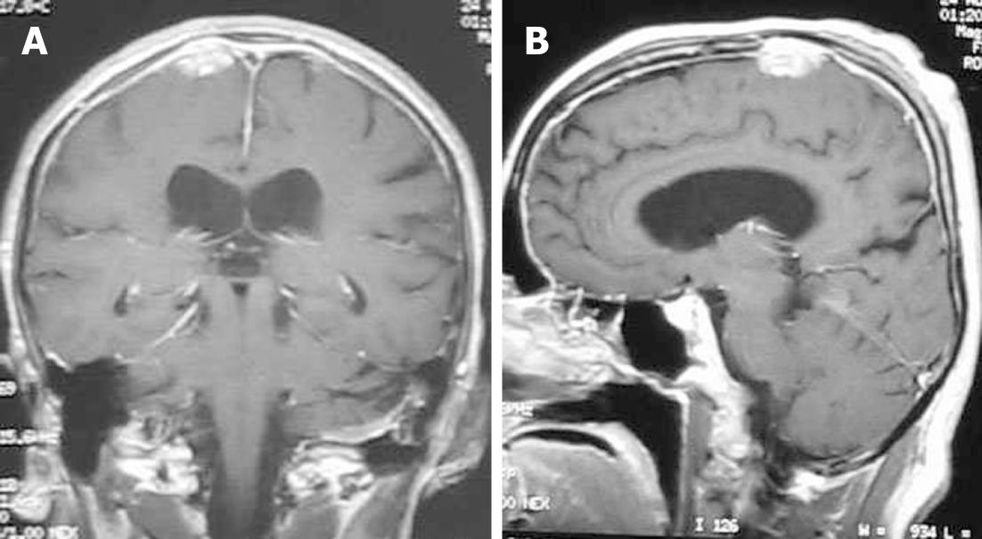
Figure 8 Neurosarcoidosis.
Coronal (A) and sagittal (B) T1-weighted magnetic resonance post-contrast images show an enhancing dural-based lesion. Also, note the enhancement of the dura along the convexity.
Figure 9 Melanocytic tumor.
A: Photomicrograph of the mass. The neoplasm contains sheets of cells, many of which contain dark brown melanin pigment (hematoxylin-eosin, × 400); B: T1-weighted magnetic resonance and post-contrast; C: T1-weighted MR images show a left intraconal mass with intermediate signal intensity that homogeneously enhances.
Figure 10 Plasma cell granuloma in a 4-year-old girl.
A: Pathologic image. The fibrosis and infiltration is composed of lymphocytes, plasmocytes and histiocytes (hematoxylin-eosin stain); B: Axial T2-weighted magnetic resonance (MR) image shows a hypointense left cerebellar lesion adjacent to the 4th ventricle with moderate peritumoral edema; C: Post-contrast T1-weighted MR image shows heterogeneous enhancement of the lesion.
- Citation: Chourmouzi D, Potsi S, Moumtzouoglou A, Papadopoulou E, Drevelegas K, Zaraboukas T, Drevelegas A. Dural lesions mimicking meningiomas: A pictorial essay. World J Radiol 2012; 4(3): 75-82
- URL: https://www.wjgnet.com/1949-8470/full/v4/i3/75.htm
- DOI: https://dx.doi.org/10.4329/wjr.v4.i3.75


