Copyright
©2012 Baishideng Publishing Group Co.
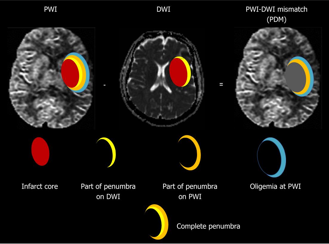
Figure 1 Modern concept of ischemic penumbra.
Early abnormality on diffusion weighted imaging (DWI) equals the infarct core plus a part of the tissue at risk (penumbra); and the perfusion deficiency on perfusion weighted imaging (PWI) includes the infarct core plus penumbra and region of benign oligemia. PWI-DWI mismatch (PDM) does not optimally define the ischemic penumbra.
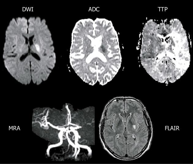
Figure 2 Main pattern of perfusion-diffusion mismatch, perfusion-weighted imaging > diffusion-weighted imaging, in a patient with acute stroke.
Extensive area of prolonged time to peak (TTP) and small diffusion-weighted imaging (DWI) lesion in deep middle cerebral artery (MCA) territory, with a complete proximal MCA occlusion on magnetic resonance angiography (MRA) (reprint from Muir KW et al Lancet Neurol 2006; 5: 755-68 with permission). ADC: Apparent diffusion coefficient; FLAIR: Fluid-attenuated inversion-recovery.
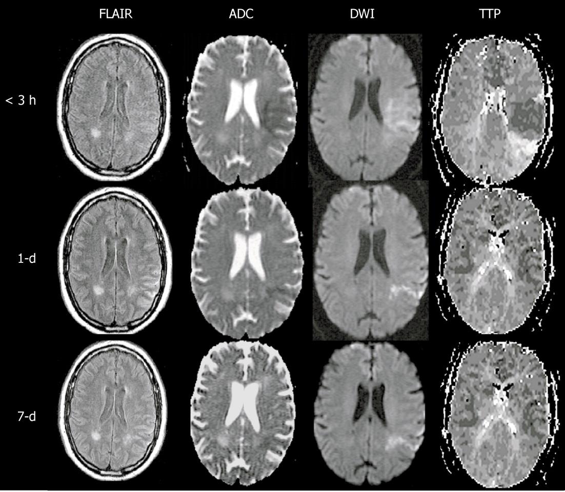
Figure 3 Main pattern of perfusion-diffusion mismatch, perfusion-weighted imaging = diffusion-weighted imaging, in a patient with acute stroke.
< 3 h: Fuzzy diffusion-weighted imaging (DWI) lesion in left middle cerebral artery territory matching an area of diminished time to peak, indicating local hyperperfusion and spontaneous recanalization had occurred prior to imaging at 3 h after onset (note the prolonged time to peak at the posterior edge of the DWI lesion, suggesting distal branch occlusion); 1-d: The next day, perfusion has essentially normalized as well as the DWI lesion, save for a narrow posterior streak, suggesting the spontaneous recanalization saved the at-risk tissue from progressing to infarction; 7-d: At day 7, there has been no return of the DWI lesion, indicating the tissue was effectively salvaged (reprint from Muir KW et al Lancet Neurol 2006; 5: 755-768 with permission). PWI: Perfusion-weighted imaging; ADC: Apparent diffusion coefficient; TTP: Time to peak; FLAIR: Fluid-attenuated inversion-recovery.
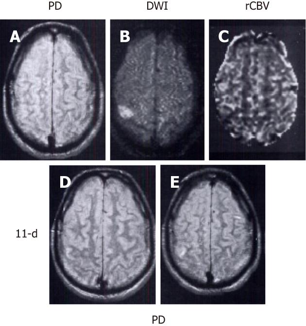
Figure 4 Main pattern of perfusion-diffusion mismatch: perfusion-weighted imaging < diffusion-weighted imaging, in a patient with acute stroke.
Patient’s left-sided weakness was partially resolved 3 h after the onset of symptoms. A: Proton density (PD) weighted fast spin-echo image shows no abnormality; B: Diffusion-weighted imaging (DWI) shows a focal cortical ischemic abnormality; C: Relative cerebral blood volume (rCBV) map demonstrates a smaller lesion with decreased rCBV compared to the same abnormality as depicted in B; D, E: Follow-up PD images acquired 11 d later show a new tiny hyperintense infarct in the area of initially observed lesion on B and C. Note that the initial DWI lesion is larger than the final infarct volume. The patient’s symptoms resolved completely after 2 d (reprint from Sorensen et al Radiology 1996; 199: 391-401 with permission).
- Citation: Chen F, Ni YC. Magnetic resonance diffusion-perfusion mismatch in acute ischemic stroke: An update. World J Radiol 2012; 4(3): 63-74
- URL: https://www.wjgnet.com/1949-8470/full/v4/i3/63.htm
- DOI: https://dx.doi.org/10.4329/wjr.v4.i3.63


