Copyright
©2012 Baishideng Publishing Group Co.
World J Radiol. Mar 28, 2012; 4(3): 115-120
Published online Mar 28, 2012. doi: 10.4329/wjr.v4.i3.115
Published online Mar 28, 2012. doi: 10.4329/wjr.v4.i3.115
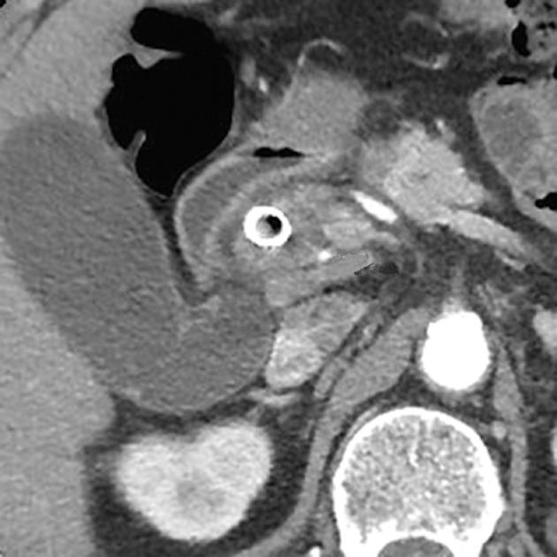
Figure 1 Computed tomography image obtained 5 mo after self-expandable metallic stent insertion showed no abnormal findings of the right hepatic artery (arrow).
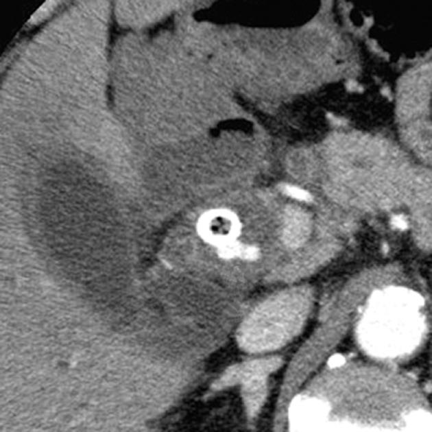
Figure 2 Computed tomography showed a small pseudoaneurysm of the right hepatic artery, measuring 9 × 6 mm, protruding into the common bile duct lumen (arrow) 9 mo after self-expandable metallic stent insertion.
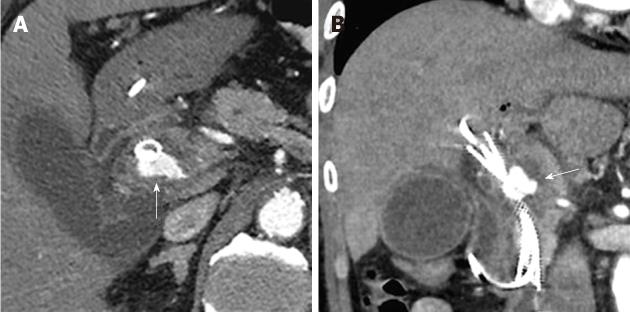
Figure 3 On the 2nd hospital day, an endoscopic naso-biliary drainage and a plastic stent were placed into the self-expandable metallic stent.
On the 6th hospital day, axial (A) and coronal (B) plane arterial phase computed tomography showed enlargement of the pseudoaneurysm (arrow).
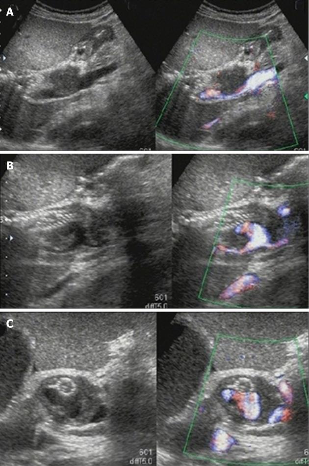
Figure 4 Color Doppler ultrasonography on the 6th hospital day.
A: Ultrasonography showed marked dilation of the common bile duct to a diameter of 24 mm and consequent extrinsic compression of the portal vein; B:The self-expandable metallic stent (SEMS) was displaced toward the liver, and hypoechoic solid components filling the space between the SEMS and bile duct and a pseudoaneurysm showing cystic growth in the lumen of the bile duct was observed; C: Only the apex of the pseudoaneurysm was in contact with the SEMS.
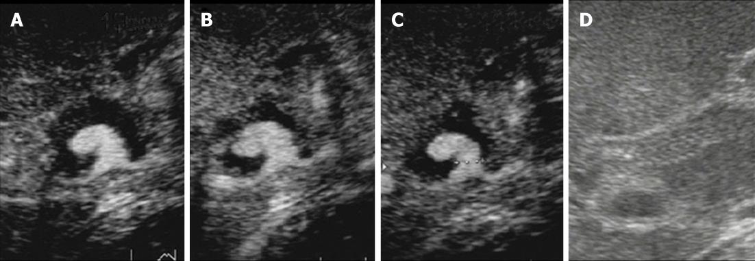
Figure 5 The pseudoaneurysm and hypoechoic solid components filling the dilated bile duct were examined on contrast-enhanced ultrasonography.
A-C: Images obtained at 15 s (A), 55 s (B) and 127 s (C) after injection of Sonazoid (0.5 mL) via a left cubital venous line showed no Sonazoid bubbles in the common bile duct other than in the pseudoaneurysm; D: Monitor B-mode ultrasonography image.
Figure 6 Transcatheter arterial embolization performed on the 12th hospital day.
A, B: On angiography, the left and right hepatic arteries were found to arise from the superior mesenteric artery, and the pseudoaneurysm was confirmed to be located in the right hepatic artery; C: One IDC coil was placed in the right hepatic artery on the distal side of the pseudoaneurysm (arrow) after the pseudoaneurysm was framed using several types of coils. Seven coils were placed in the right hepatic artery on the distal and proximal sides of the pseudoaneurysm using the isolation method.
- Citation: Watanabe M, Shiozawa K, Mimura T, Ito K, Kamata I, Kishimoto Y, Momiyama K, Igarashi Y, Sumino Y. Hepatic artery pseudoaneurysm after endoscopic biliary stenting for bile duct cancer. World J Radiol 2012; 4(3): 115-120
- URL: https://www.wjgnet.com/1949-8470/full/v4/i3/115.htm
- DOI: https://dx.doi.org/10.4329/wjr.v4.i3.115


