Copyright
©2012 Baishideng Publishing Group Co.
World J Radiol. Dec 28, 2012; 4(12): 462-468
Published online Dec 28, 2012. doi: 10.4329/wjr.v4.i12.462
Published online Dec 28, 2012. doi: 10.4329/wjr.v4.i12.462
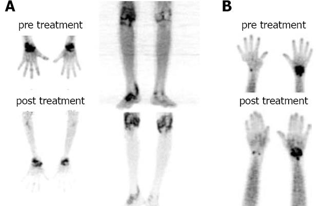
Figure 1 Images of response assessment in rheumatoid arthritis.
The patient on the left side was a responder and right side was a non-responder. A: A good response to the hydroxyquinone, methotrexate and prednisolone treatment (at 6 wk) on follow up scan. The pre treatment maximum standardized uptake value (SUVmax) values were 10.6 in right wrist which fell to 4.0 on post treatment scan; B: An example of non-responder. The pre treatment SUVmax value was 2.9 while that of post treatment with hydroxyquinolone, methotrexate and prednisolone was 4.3. The similarity of the uptake in pre and post treatment scan made it difficult to interpret progression on visual basis. The patient had no symptomatic relief clinically.
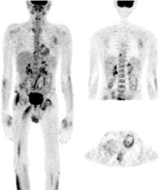
Figure 2 The asymmetrical fluorodeoxyglucose uptake pattern in a newly diagnosed patient with ankylosing spondylitis.
The asymmetrical heterogeneous fluorodeoxyglucose (FDG) uptake is noted right sternoclavicular, left hip and a solitary facetal joint in dorsal spine. High to intense FDG uptake is seen in soft tissue of right thigh (probably fascia lata) and right lower limb muscles. Even one could notice the soft tissue FDG activity in tendons of small joints of hands.
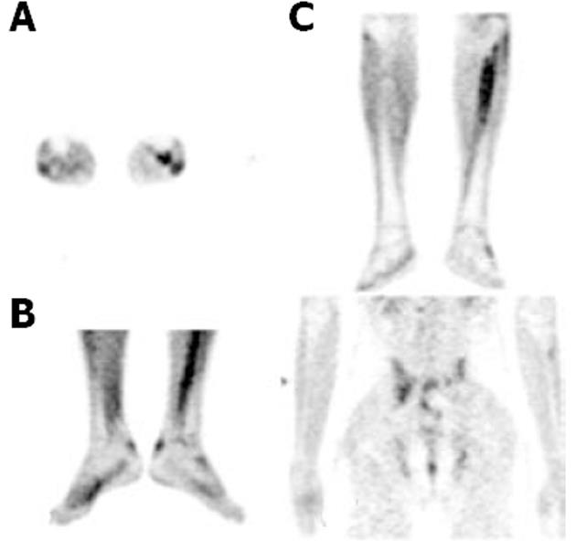
Figure 3 Soft tissue uptake in patient with seronegative spondyloarthropathy.
A: The fibrous tissue uptake in the interosseous membrane of left leg; B: The bilateral calcaneal tendonal tracer activity; C: Fluorodeoxyglucose (FDG)-positron emission tomography in another patient with ankylosing spondylitis demonstraing the heterogeneous FDG uptake in the bilateral sacroiliac joints.
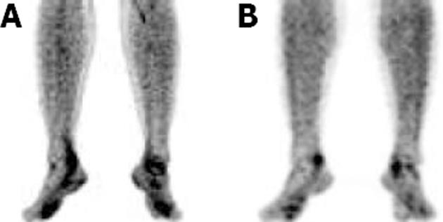
Figure 4 Assessment of treatment response in a patient of psoriatic arthritis.
A: Pre treatment; B: Post treatment. The fluorodeoxyglucose-positron emission tomography lower-limb scan of a patient with psoriatic arthritis. Rest of the whole body survey was unremarkable. Same patient was followed and a response evaluation scan was performed 6 wk after specific treatment for psoriatic arthropathy. There was significant fall in maximum standardized uptake value values correlating to the clinical improvement in the patient.
- Citation: Vijayant V, Sarma M, Aurangabadkar H, Bichile L, Basu S. Potential of 18 F-FDG-PET as a valuable adjunct to clinical and response assessment in rheumatoid arthritis and seronegative spondyloarthropathies. World J Radiol 2012; 4(12): 462-468
- URL: https://www.wjgnet.com/1949-8470/full/v4/i12/462.htm
- DOI: https://dx.doi.org/10.4329/wjr.v4.i12.462


