Copyright
©2012 Baishideng Publishing Group Co.
World J Radiol. Dec 28, 2012; 4(12): 455-461
Published online Dec 28, 2012. doi: 10.4329/wjr.v4.i12.455
Published online Dec 28, 2012. doi: 10.4329/wjr.v4.i12.455
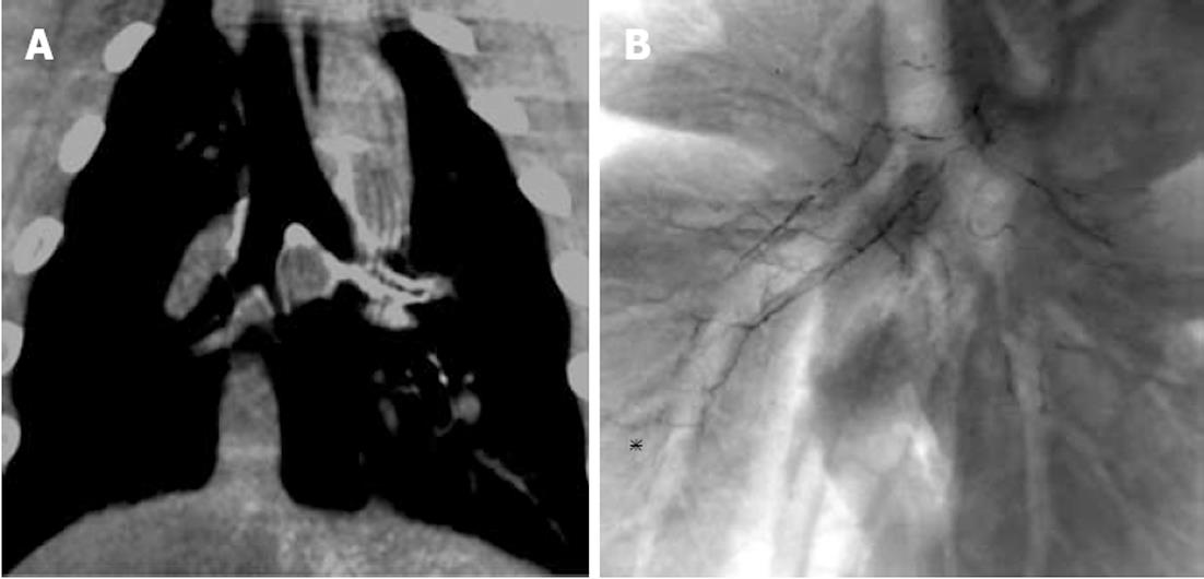
Figure 1 Embolization of right bronchial artery with n-butyl cyanoacryrate lipiodol was conducted.
Two days after bronchial artery embolization (BAE) of bilateral bronchial arteries, chest computed tomography showed accumulation of n-butyl cyanoacryrate lipiodol (NBCA-Lp) alongside the bronchial trees (A). Chest radiography of the sacrificed lung two days after BAE with NBCA-Lp showed accumulation of NBCA-Lp that corresponded to the bilateral branch arteries of the main bronchus, lobar bronchus, and segmental (asterisk) bronchi (B).
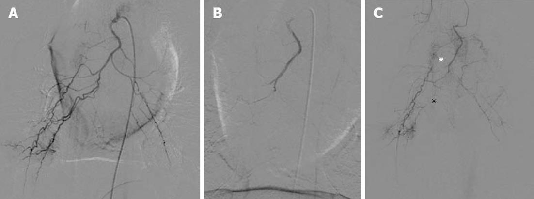
Figure 2 Common tract bronchial arteriography before (A) and immediately after (B) embolization with gelatin sponge particles.
Two days after (C), common tract bronchial arteriography showed overt stenosis (black asterisk) and occlusion (white asterisk) of the bronchial branch arteries.
Figure 3 Microscopic study.
A: The bronchial branch artery with embolization of n-butyl cyanoacrylate lipiodol (NBCA-Lp) shows that the bubble-like space corresponds to NBCA-Lp (asterisks) containing red thrombus (arrowhead), with stripping of endothelial cells (arrow) and infiltration of the inflammatory cells (hematoxylin eosin stain × 100); B: The bronchial branch artery with embolization of gelatin sponge particles (GSP) shows amorphous substance corresponding to GSP (arrow) surrounded by red thrombus with infiltration of inflammatory cells (asterisks) (hematoxylin eosin stain × 100); C: The bronchial wall revealed the bronchial branch artery (arrows) embolized with NBCA-Lp outside the cartilage and the patency of numerous fine networks (asterisks) in the submucosal and cartilage layers (hematoxylin eosin stain × 100).
- Citation: Tanaka T, Kawai N, Sato M, Ikoma A, Nakata K, Sanda H, Minamiguchi H, Nakai M, Sonomura T, Mori I. Safety of bronchial arterial embolization with n-butyl cyanoacrylate in a swine model. World J Radiol 2012; 4(12): 455-461
- URL: https://www.wjgnet.com/1949-8470/full/v4/i12/455.htm
- DOI: https://dx.doi.org/10.4329/wjr.v4.i12.455


