Copyright
©2011 Baishideng Publishing Group Co.
World J Radiol. Sep 28, 2011; 3(9): 224-232
Published online Sep 28, 2011. doi: 10.4329/wjr.v3.i9.224
Published online Sep 28, 2011. doi: 10.4329/wjr.v3.i9.224
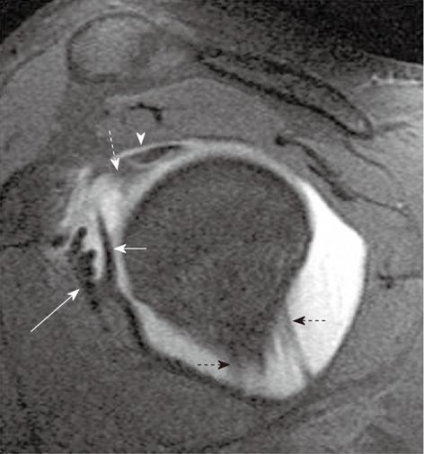
Figure 1 Normal T1-weighted TSE fat-saturated axial magnetic resonance arthrogram image.
The anterior and posterior labrum appears as triangular hypointense structures (straight arrows). Normal middle glenohumeral ligament has been shown with an arrowhead. Note the long head of biceps tendon in the bicipital groove and extension of joint fluid around the tendon (dashed arrow).
Figure 2 Axial T1-weighted fat-saturated magnetic resonance arthrogram image shows normal superior glenohumeral ligament (straight arrow) running parallel to the coracoid concavity and the long head of biceps tendon (dashed arrow).
Figure 3 Axial T1-weighted fat-saturated magnetic resonance arthrogram image shows normal anterior and posterior bands of inferior glenohumeral ligament (straight arrows).
Posterior labrum is seen as normal hypointense structure (dashed arrow), anterior labrum is congenitally absent in this patient.
Figure 4 Oblique sagittal T1-weighted fat-saturated magnetic resonance arthrogram image shows normal superior glenohumeral ligament (white dashed arrow), inferior to the intra-articular long head of biceps tendon (arrowhead).
The middle glenohumeral ligament is seen as a long hypointense band (short straight arrow) medial to the subscapularis tendon (long straight arrow). Anterior and posterior bands of inferior glenohumeral ligament are shown with black dashed arrows.
Figure 5 Magnetic resonance arthrographic axial T1-weighted fat-saturated images showing different types of attachment of anterior joint capsule.
A: Type I; B: Type II; C: Type III (arrows).
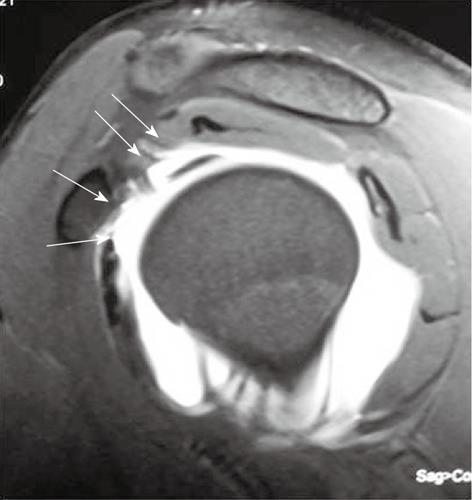
Figure 6 Oblique sagittal T1-weighted fat-saturated magnetic resonance arthrogram image shows normal rotator cuff interval (arrows).
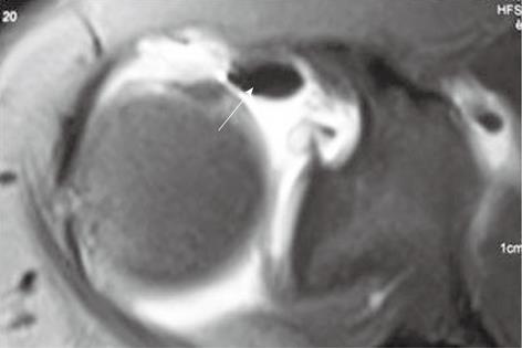
Figure 7 Axial T1-weighted fat-saturated magnetic resonance arthrogram image shows artifact due to inadvertent injection of air into the joint cavity.
The air appears as hypointense structure lying in nondependent areas (arrow), which helps differentiate it from loose bodies.
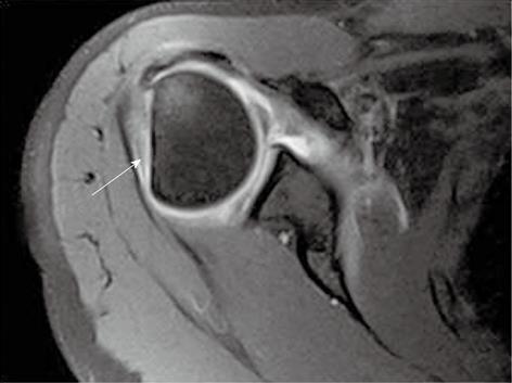
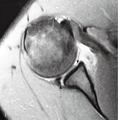
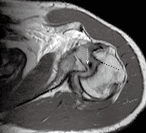
Figure 8 Axial T1-weighted TSE fat-suppressed magnetic resonance arthrogram image shows bony defect involving posterosuperior humeral head (Hill-Sachs lesion) (arrow).
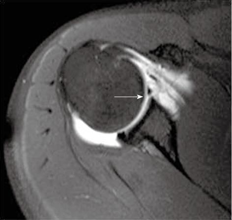
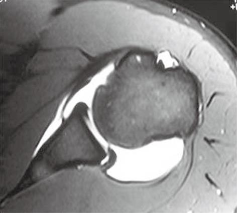
Figure 9 Axial T1-weighted TSE fat-suppressed magnetic resonance arthrogram image shows detached anteroinferior labrum from the glenoid margin; classic soft tissue Bankart lesion.
Contrast within the joint is seen to traverse the gap between the detached labrum and the glenoid margin (arrow).
Figure 10 Axial T1-weighted TSE fat-suppressed magnetic resonance arthrogram image shows anteroinferior labral tear with bony glenoid injury shown with an arrow (Bony Bankart lesion).
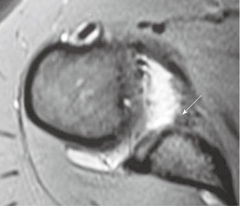
Figure 11 Perthes lesion.
Oblique axial T2-weighted TSE image of the shoulder joint with the arm in abduction and external rotation location shows tear of the anteroinferior labrum (arrow) with intact periosteum, suggesting Perthes lesion.
Figure 12 Anterior labroligamentous periosteal sleeve avulsion lesion in a patient with recurrent anterior shoulder dislocation.
A: Axial T2-weighted gradient-echo image of the right shoulder reveals irregular contour of the anteroinferior labrum and hypointense soft tissue lying along the scapular neck (arrow); B: On magnetic resonance arthrographic axial T1-weighted fat-saturated image the avulsed labroligamentous tissue is seen displaced medially along the scapular neck (arrow).
Figure 13 Glenolabral articular disruption lesion and posterior labral tear in a patient with multidirectional instability.
Axial proton density fat-suppressed image reveals absence of the anteroinferior labrum with tear of the adjacent articular cartilage (straight arrow). Also associated is a tear involving the posterior labrum, seen as interposition of fluid between the posterior labrum and the posterior glenoid margin (dashed arrow).
Figure 14 Humeral avulsion of anterior glenohumeral ligament in chronic anterior instability.
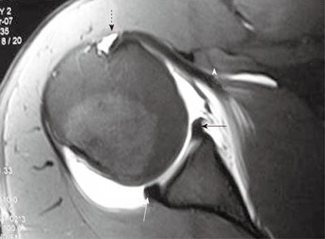
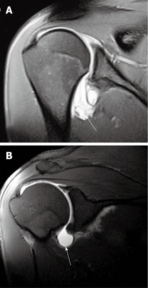
Coronal T1-weighted TSE fat-suppressed magnetic resonance arthrogram image reveals the ‘J’ shape (arrow) of the axillary pouch (A), compared to the ‘U’ shape (arrow) in a normal individual (B).
Figure 15 Glenolabral articular disruption lesion.
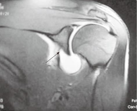
Coronal T1-weighted TSE fat-suppressed magnetic resonance arthrogram image reveals avulsion of the glenoid attachment of the anterior band of the inferior glenohumeral ligament (arrow).
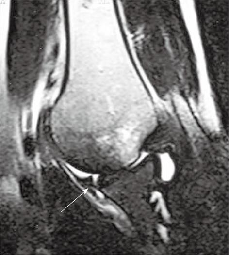
Figure 16 Coronal TSE T2-weighted image of the right shoulder in a patient with acute dislocation reveals full thickness tear of the supraspinatus tendon with proximal retraction of the muscle (arrow).
Figure 17 Buford complex.
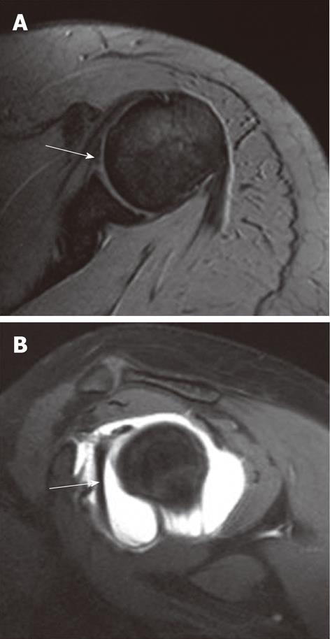
A: Axial T2-weighted gradient-echo image reveals absent anterior labrum and a thick hypointense structure lying anteriorly (arrow) which can be mistaken for a torn labrum; B: Oblique sagittal T1-weighted TSE fat-saturated magnetic resonance arthrogram image reveals a thick cord-like middle glenohumeral ligament (arrow) having a higher glenoid attachment, close to 12 o’clock location.
Figure 18 Reverse Hill-Sachs and reverse Bankart lesion in a case of posterior instability.
T1-weighted TSE axial magnetic resonance image reveals hemarthrosis, posterior glenohumeral dislocation and reverse Hill-Sachs lesion (straight arrow). There is associated posterior labral tear (reverse Bankart lesion), shown with a dashed arrow.
Figure 19 Posterior labral tear.
Axial T1-weighted TSE fat-suppressed magnetic resonance arthrogram image reveals the undisplaced posterior labral tear (arrow). The anterior joint capsule attachment is placed medially along scapular neck (normal variation).
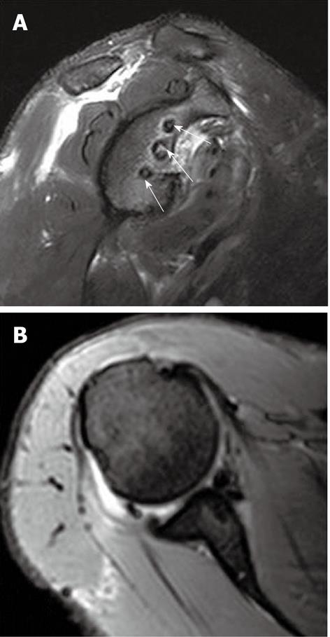
Figure 20 Normal post-operative appearance after arthroscopic suture-anchor repair of Bankart lesion.
Oblique sagittal (A) and axial (B) T2-weighted TSE fat-suppressed image reveals the three suture-anchors in place (arrows). No fluid is seen between the labral margin and the opposed labrum and joint capsule.
- Citation: Jana M, Gamanagatti S. Magnetic resonance imaging in glenohumeral instability. World J Radiol 2011; 3(9): 224-232
- URL: https://www.wjgnet.com/1949-8470/full/v3/i9/224.htm
- DOI: https://dx.doi.org/10.4329/wjr.v3.i9.224


