Copyright
©2011 Baishideng Publishing Group Co.
World J Radiol. Jul 28, 2011; 3(7): 182-187
Published online Jul 28, 2011. doi: 10.4329/wjr.v3.i7.182
Published online Jul 28, 2011. doi: 10.4329/wjr.v3.i7.182
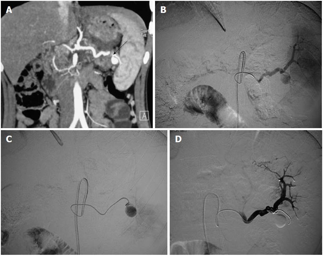
Figure 1 Splenic artery aneurysm in a 21-year-old asymptomatic female.
A: Coronal maximum intensity projection image of computed tomography angiogram reveals a pseudoaneurysm from the distal splenic artery at the hilum with a narrow neck (arrow); B, C: Digital subtraction angiography images reveal the pseudoaneurysm; D: After selective catheterization, the aneurysm was treated by coiling which resulted in complete occlusion in the aneurysm sac; visualized in the post-procedure image.
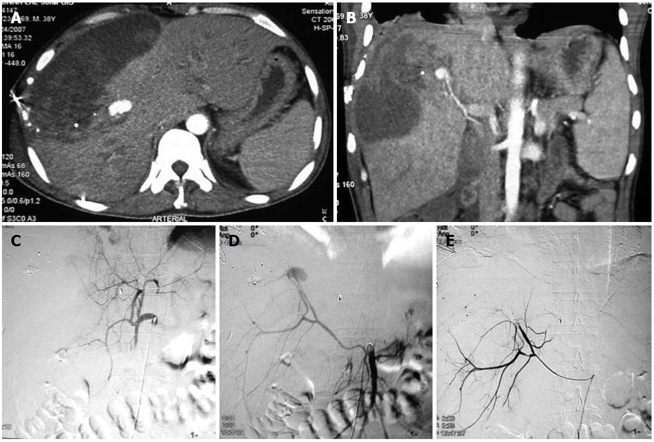
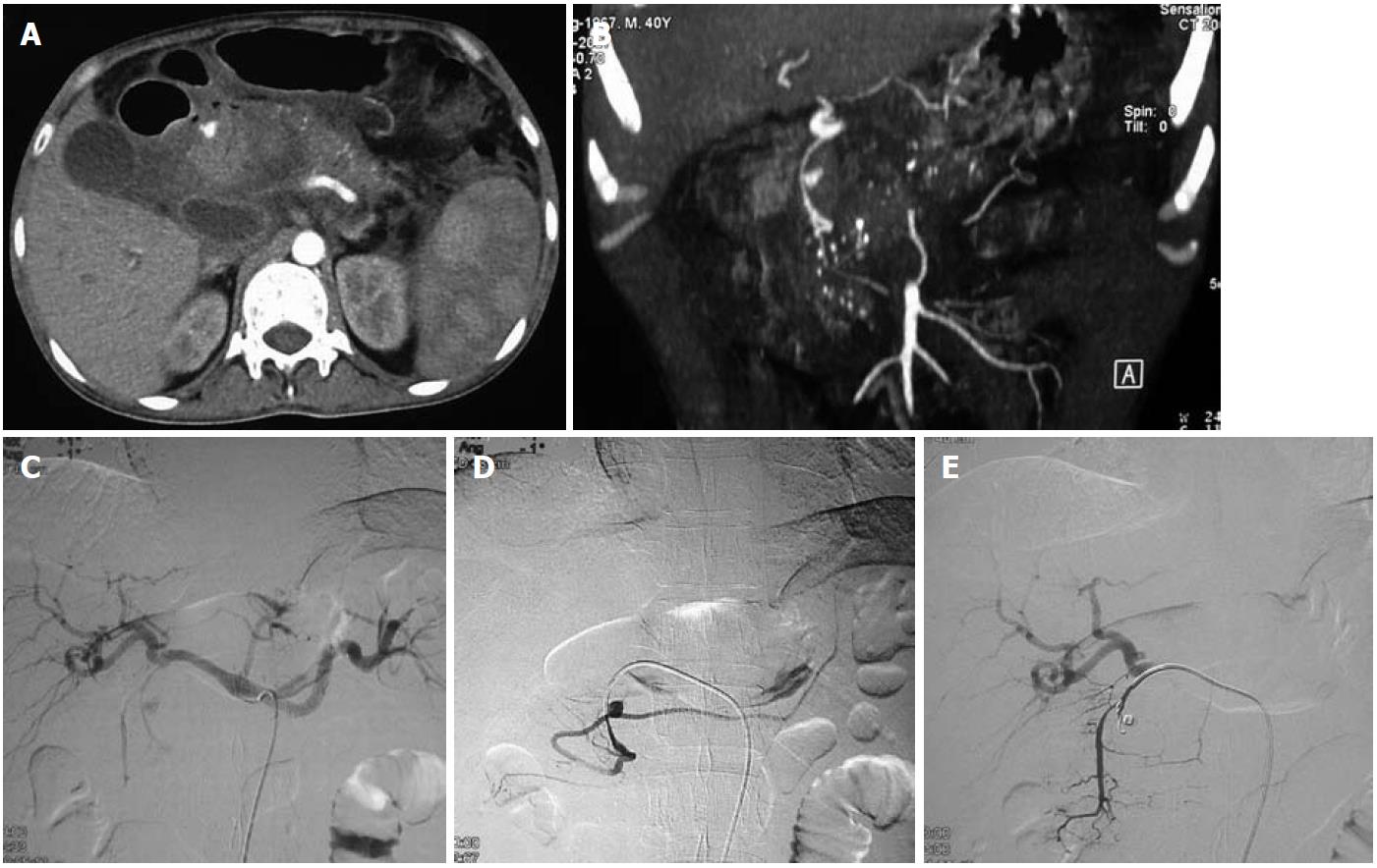
Figure 2 Traumatic pseudoaneurysm of a replaced right hepatic artery.
A 38-year-old male presented with hypotension and falling hematocrit after a road traffic accident. A, B: Axial contrast-enhanced computed tomography of the abdomen (A) and coronal MIP image (B) revealed a large pseudoaneurysm (arrows) in the right lobe of the liver and a large hematoma in the liver parenchyma extending to the subcapsular location; C: DSA image after selective catheterization of the celiac trunk failed to reveal any aneurysm; D: Selective superior mesenteric artery (SMA) catheterization revealed a replaced right hepatic artery arising from the SMA and a pseudoaneurysm arising from it; E: Treated with coil embolization.
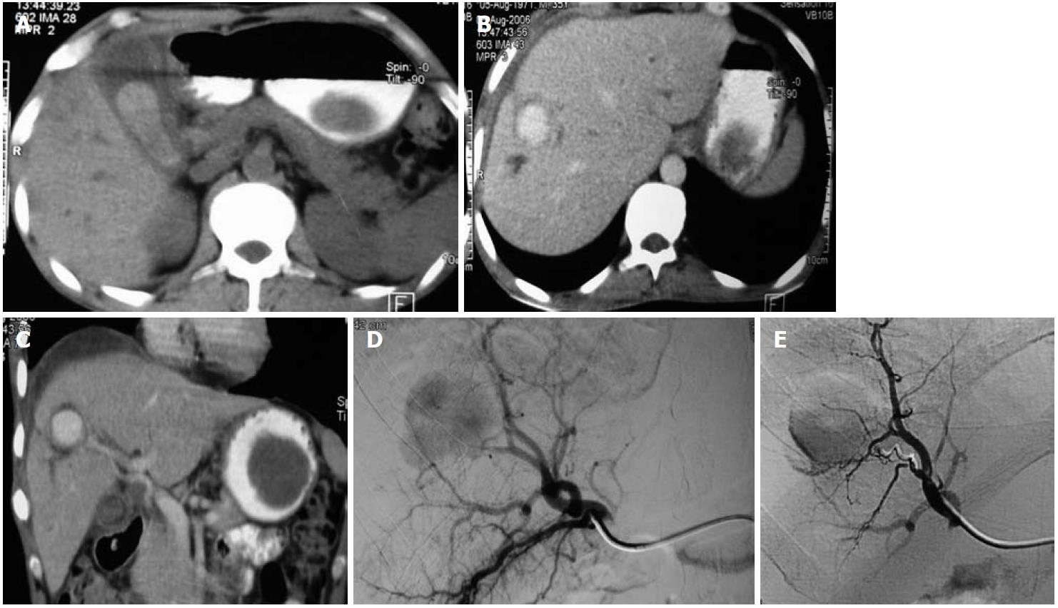
Figure 3 Traumatic right hepatic artery aneurysm.
A 45-year-old male presented 2 mo after a road traffic accident with melena. A: Abdominal non-contrast computerized tomography revealed a hyperdense hematoma in the gall bladder lumen; B, C: Abdominal contrast-enhanced computed tomography axial (B) and coronal reformatted images (C) revealed a large pseudoaneurysm of the right hepatic artery (arrows); D: Selective catheter angiogram of the hepatic artery revealed the large pseudoaneurysm; E: Successful treatment by coil embolization (e).
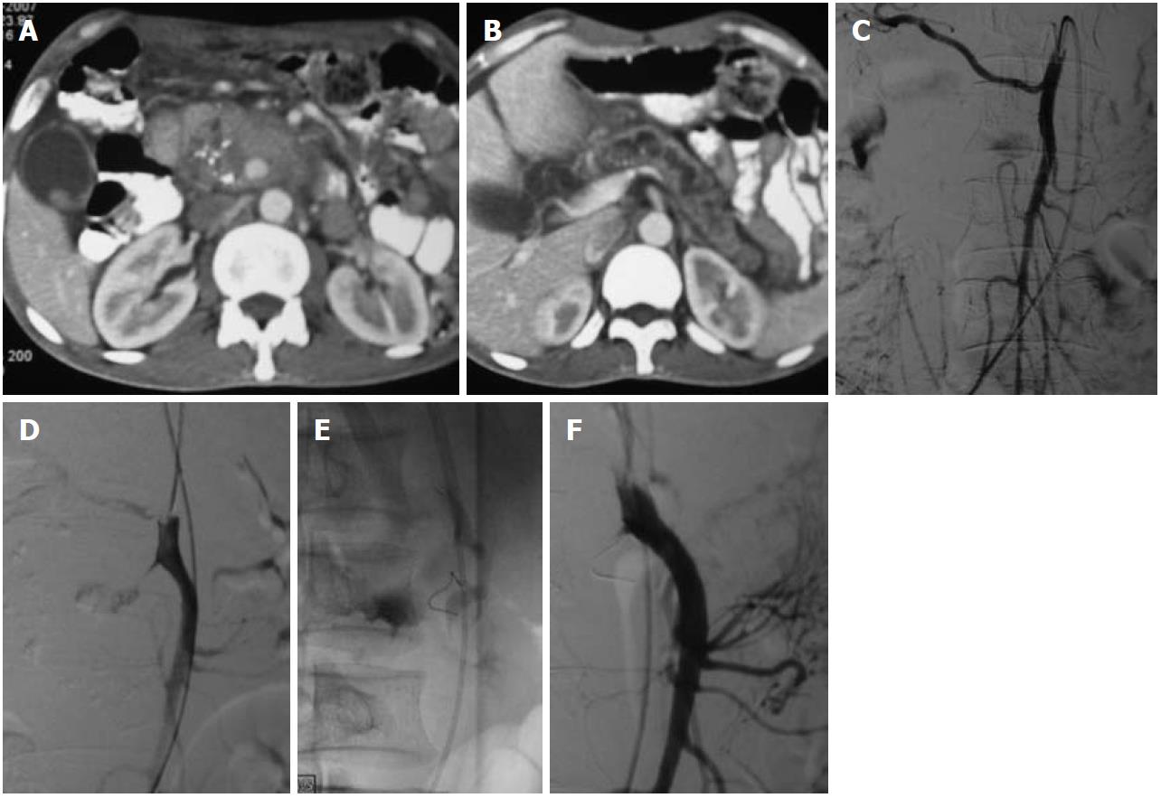
Figure 4 Coil embolization in an superior mesenteric artery aneurysm in a patient with chronic calcific pancreatitis.
A, B: Axial contrast-enhanced computed tomography of the abdomen revealed dilated main pancreatic duct and coarse calcification in the head of the pancreas, a large partially thrombosed pseudoaneurysm was apparent as a contrast filled globular structure in the head; C, D: Selective abdominal angiography of the superior mesenteric artery revealed the jet of injected contrast into the pseudoaneurysm cavity (D); E, F: Coil embolization was performed to occlude the neck (E) which resulted in complete occlusion and non-filling of the aneurysm (F).
Figure 5 Endovascular management of a gastroduodenal artery aneurysm secondary to chronic calcific pancreatitis.
A, B: Axial image (A) and coronal reformatted image (B) of a computed tomography angiogram reveal features of acute on chronic calcific pancreatitis and a small saccular pseudoaneurysm of the gastroduodenal artery; C, D: Selective angiogram of the celiac axis (C) and gastroduodenal artery (D) reveal the filling of the aneurysm from the main trunk; E: Coil embolization was performed to fill the aneurysm cavity and cause complete occlusion.
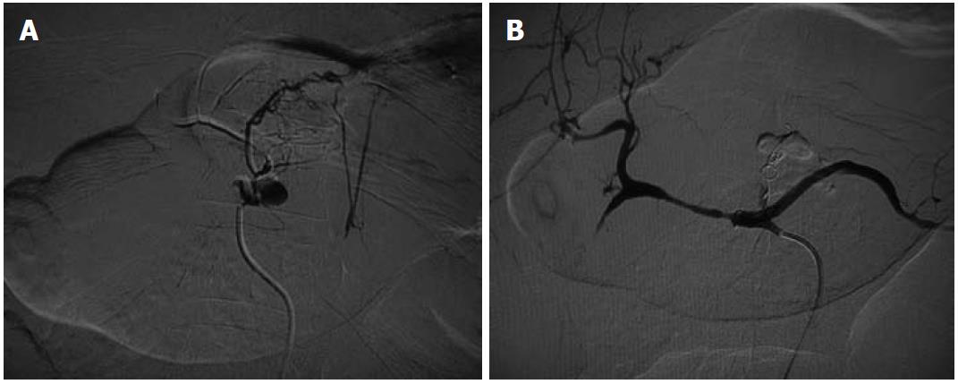
Figure 6 Endovascular coil embolization of a left gastric artery causing upper gastrointestinal hemorrhage.
A: Selective angiogram of the left gastric artery shows the large saccular aneurysm; B: Selective angiogram of the celiac axis after coil embolization revealed complete non-opacification of the left gastric artery aneurysm.
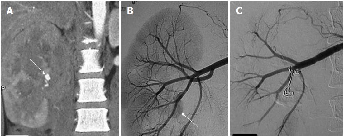
Figure 7 Traumatic right renal pseudoaneurysm.
A: Coronal maximum intensity projection computed tomography angiographic image reveals a large renal midpolar laceration and a pseudoaneurysm (arrow); B: Selective angiogram of the main right renal artery reveals a pseudoaneurysm (arrow) arising from one of the posterior branches; C: Successfully occluded using coil.
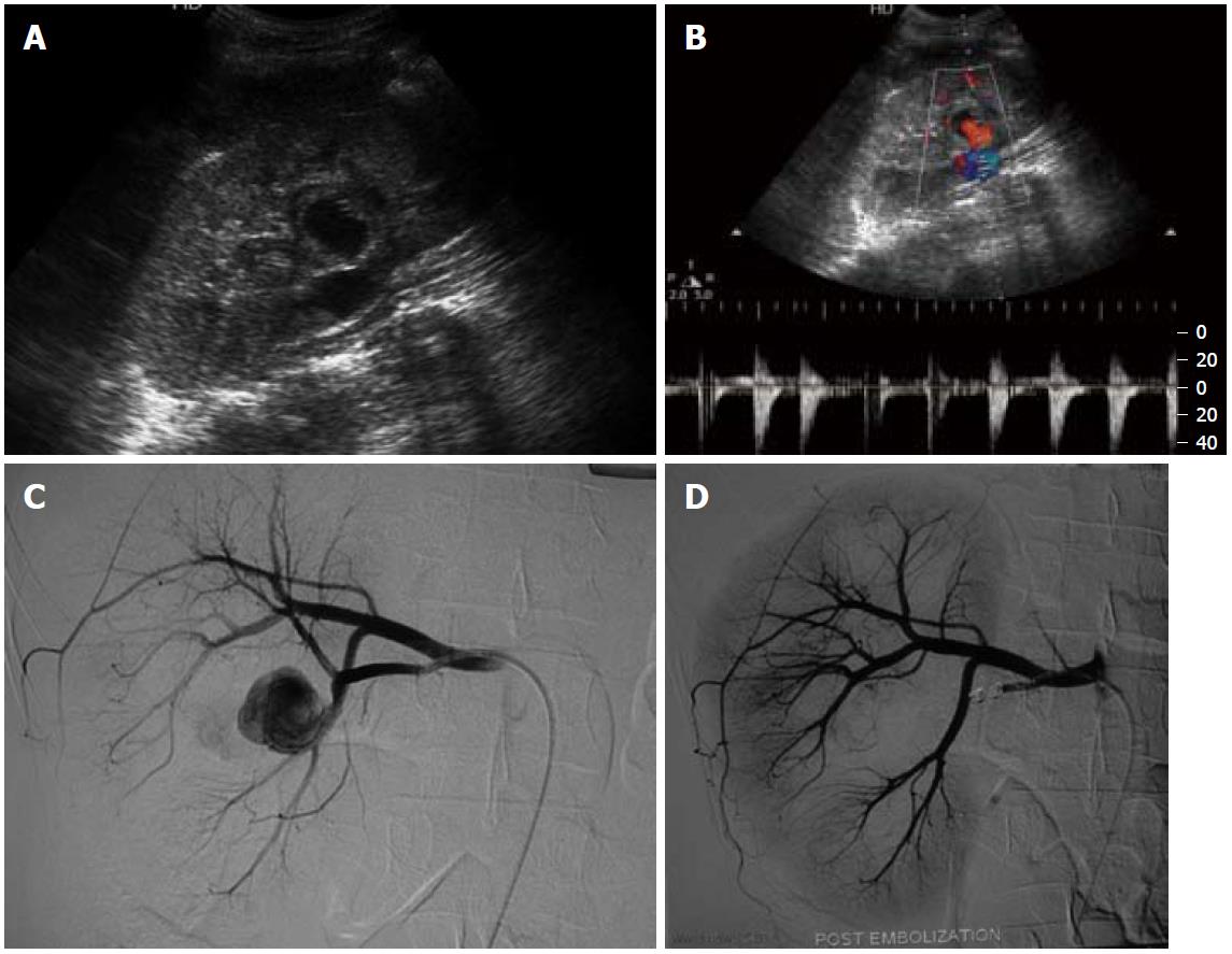
Figure 8 Traumatic right renal artery pseudoaneurysm.
A, B: Grey scale ultrasound (A) and Doppler ultrasound (B) of the right kidney reveal a pseudoaneurysm in the midpolar region; C, D: Selective angiogram (C) of the right renal artery shows the pseudoaneurysm which was successfully coil-embolized (D).
- Citation: Jana M, Gamanagatti S, Mukund A, Paul S, Gupta P, Garg P, Chattopadhyay TK, Sahni P. Endovascular management in abdominal visceral arterial aneurysms: A pictorial essay. World J Radiol 2011; 3(7): 182-187
- URL: https://www.wjgnet.com/1949-8470/full/v3/i7/182.htm
- DOI: https://dx.doi.org/10.4329/wjr.v3.i7.182


