Copyright
©2011 Baishideng Publishing Group Co.
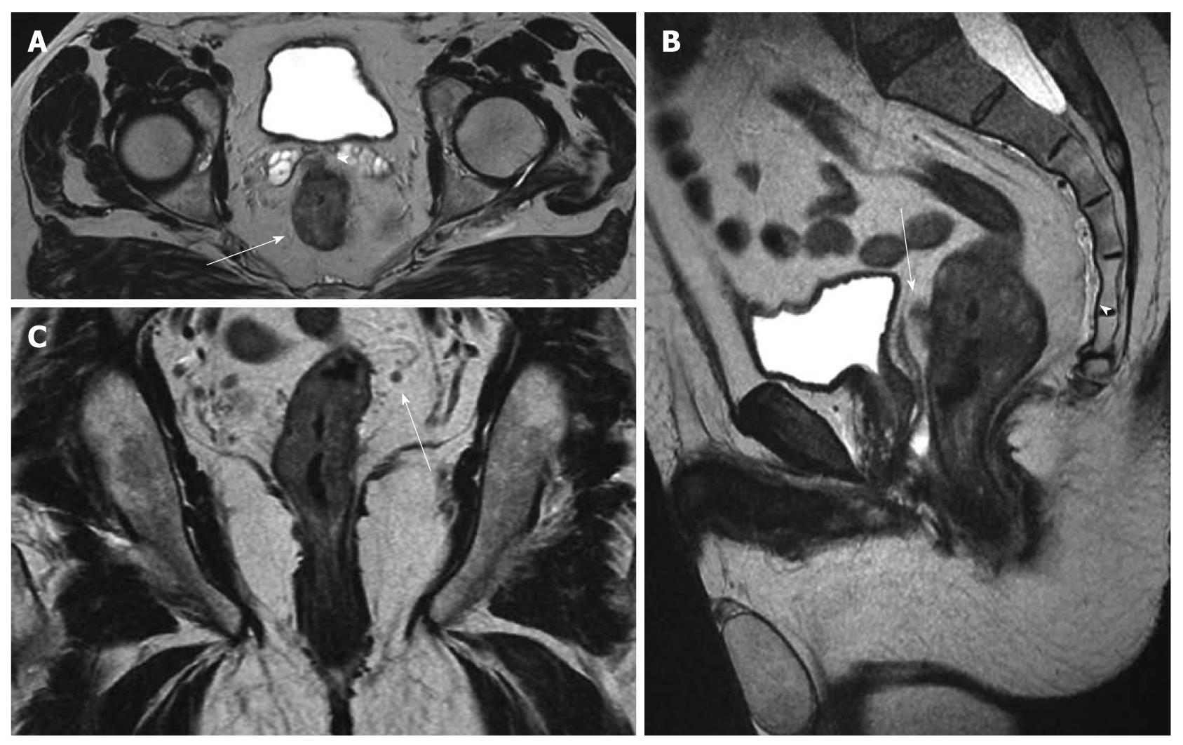
Figure 1 Magnetic resonance imaging staging of rectal cancer before chemoradiation: T3, N+.
Pathology result: T3, N1. A: Axial T2w image shows the circumferential tumor (arrow) and the extramural spread anteriorly (arrowhead) close to the seminal vesicles; B: In the sagittal T2w image, the anterior extramural spread (arrow) can be also recognized close to the mesorectal fascia (thin vertical hypointense line posterior to the bladder). The presacral fascia can also be appreciated (arrowhead) continuing inferiorly as the rectosacral fascia; C: In the coronal T2w image, a small mesorectal lymph node (arrow) is seen.
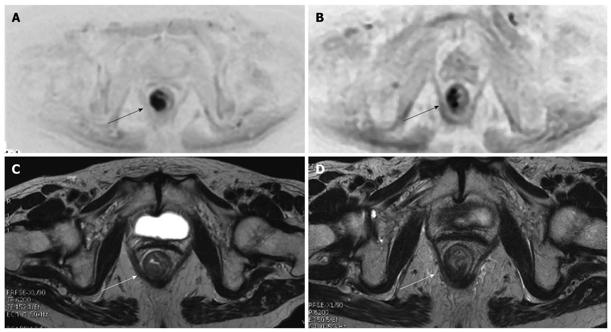
Figure 2 Diffuse weighted imaging and cancer (arrows) of the lower rectum.
At initial MR staging T3N1. After neoadjuvant therapy, MR restage was T3N0. At pathology: T3N1 (only one small metastatic mesorectal lymph node). Diffusion weighted imaging (DWI) (A) and axial T2w (B) images show the tumor (arrows) before chemoradiotherapy. The DWI image allows for better recognition of the lesion. DWI (C) and axial T2w (D) images after chemoradiotherapy show a reduction in the dimensions of the lesion (arrows).
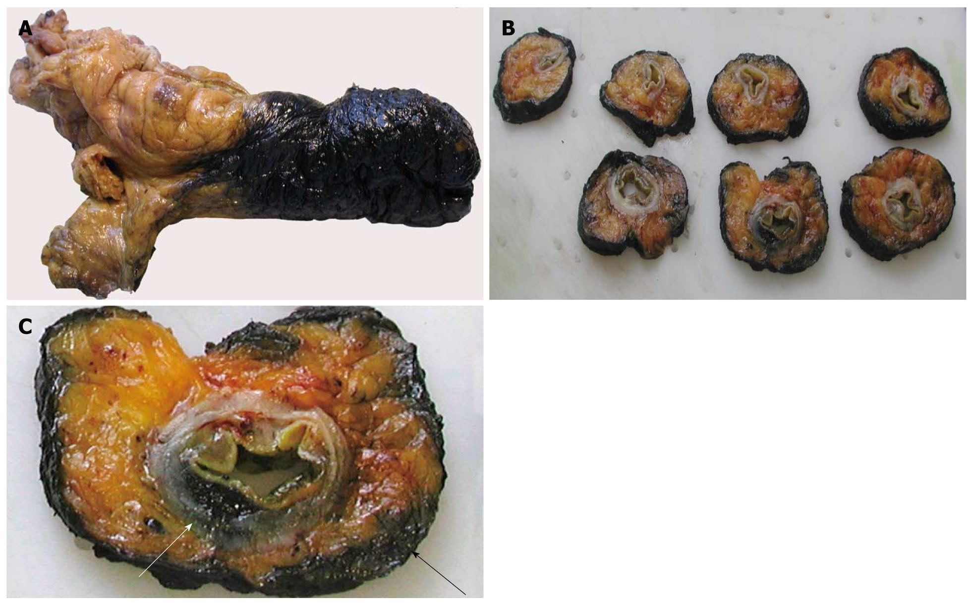
Figure 3 Pathological evaluation of the circumferential resection margin (CRM).
A: The excised rectum is dipped in ink; B: Serial sections including the tumor are taken through the entire rectum; C: One of the sections shows the tumor’s leading edge (white arrow) and its relation with the CRM (black arrow).
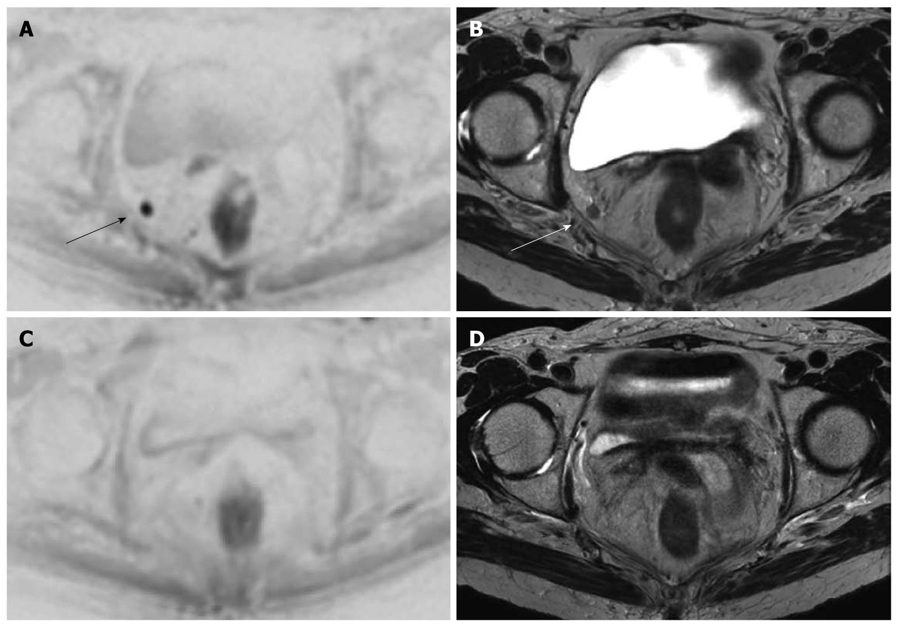
Figure 4 Same case as Figure 2.
An enlarged right obturator lymph node is suspected in this patient with a lower third rectal cancer. Diffusion weighted imaging (DWI) (A) and axial T2w (B) images show the enlarged obturator lymph node nicely (arrows). DWI (C) and axial T2w (D) images following chemoradiotherapy show that the lymph node has disappeared
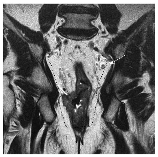
Figure 5 This case highlights the importance of the coronal plane in the assessment of the extension of the T4 lesion across the right levator complex into the right ischiorectal fossa, also shown (arrow) an enlarged and suspect lymph node.
In this case, the surgeon may choose a modified APR resection (black dotted line).
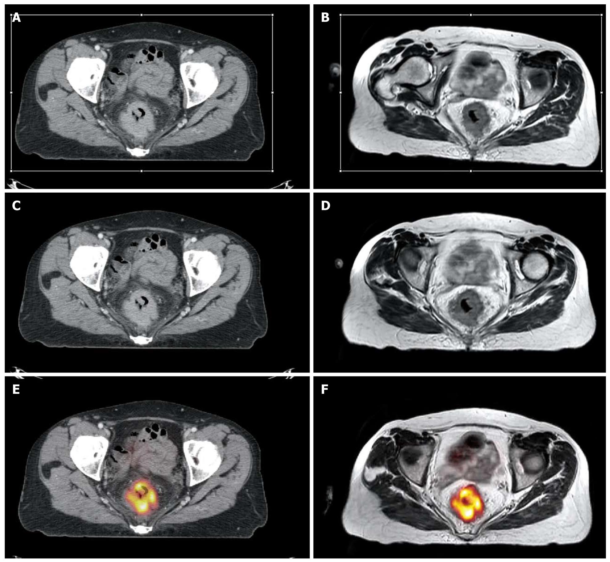
Figure 6 Multimodal image registration can be performed to improve staging and radiotherapy planning of rectal cancer.
Axial contrast enhanced computed tomography (CT) at the portal phase (A) and rigid registration of the axial T2w magnetic resonance (MR) image (B). Rigid registration reduces the misalignment between the anatomical structures in the two modalities. A, B: Volume selection (rectangular white thin line) for non-rigid registration is performed; C, D: Non-rigid registration compensates for the more complex deformations due to the different acquisition setting; E, F: Following non rigid registration, MR can be fused with the positron emission tomography (PET) image (F) and the result is super-imposable on the PET-CT image (E).
- Citation: Bellows CF, Jaffe B, Bacigalupo L, Pucciarelli S, Gagliardi G. Clinical significance of magnetic resonance imaging findings in rectal cancer. World J Radiol 2011; 3(4): 92-104
- URL: https://www.wjgnet.com/1949-8470/full/v3/i4/92.htm
- DOI: https://dx.doi.org/10.4329/wjr.v3.i4.92


