Copyright
©2011 Baishideng Publishing Group Co.
World J Radiol. Dec 28, 2011; 3(12): 289-297
Published online Dec 28, 2011. doi: 10.4329/wjr.v3.i12.289
Published online Dec 28, 2011. doi: 10.4329/wjr.v3.i12.289
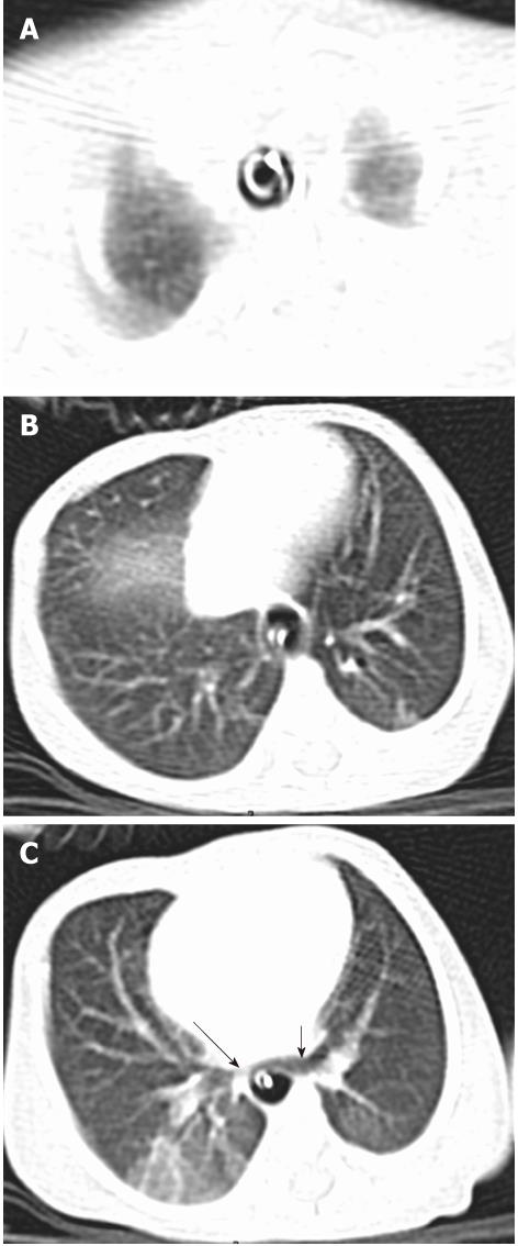
Figure 1 Tracheal agenesis type-III.
A, B: Axial images in the lung window show the presence of dilated esophagus and tube in situ. There is no separate lumen for trachea; C: Caudally, right (long arrow) and left main bronchi (short arrow) arise independently from the thoracic esophagus.
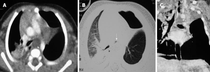
Figure 2 Foregut duplication cyst.
Axial images in the mediastinal (A) and lung (B) window and coronal multiplanar reformatted images (C) showing a fluid-attenuating lesion (long arrows) in the mediastinum compressing the left main bronchus (short arrow) with hyperinflation of left lower lobe. Hydropneumothorax in the left side was due to post-surgical change.
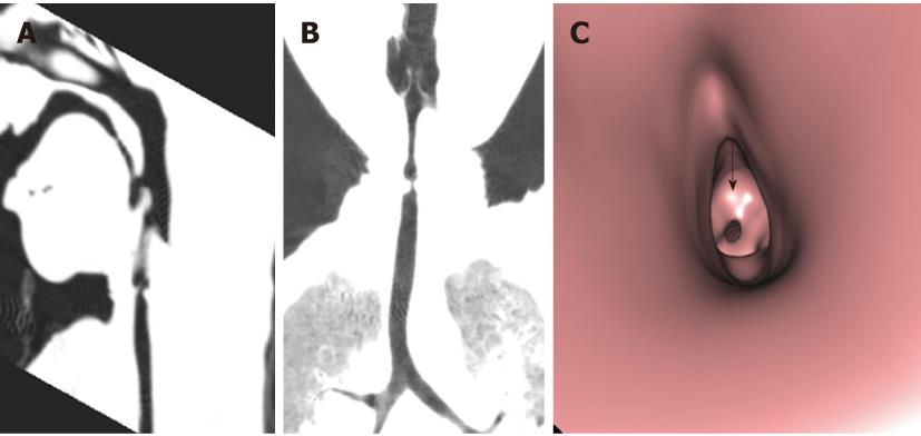
Figure 3 Congenital subglottic web.
A, B: Sagittal multiplanar reformatted images (A) and coronal minimal intensity projection images (B) show the short segment and circumferential narrowing of the subglottic region; C: Virtual bronchoscopy image shows the narrowing to be annular and is located below the vocal cords (arrow).
Figure 4 Subglottic stenosis Sagittal (A) and coronal (B) minimal intensity projection images and sagittal multiplanar reformatted images images (C) show segmental narrowing of the subglottic trachea (arrow).
Virtual bronchoscopy images from the proximal (D) and distal perspective(E) show the glottis (black arrow) and the subglottic tubular narrowing (red arrow).
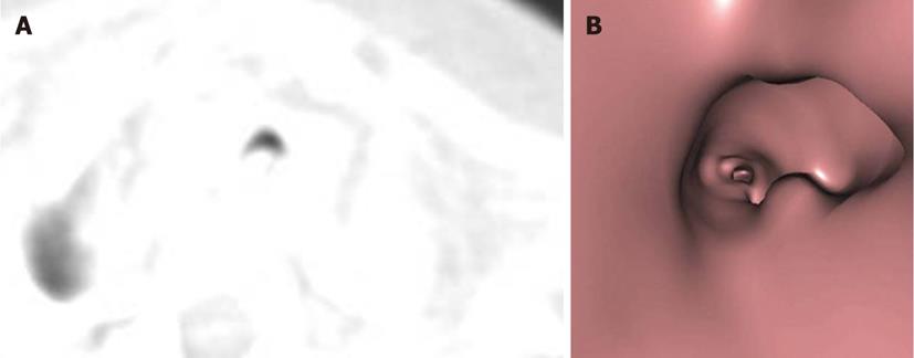
Figure 5 Tracheomalacia.
Axial image (A) in the lung window and virtual bronchoscopy image (B) shows widening of the tracheal ‘C’- cartilage and decreased antero-posterior dimension of the trachea.
Figure 6 Tracheo-esophageal fistula ‘H’- type.
Axial image in the mediastinal (A) and lung window (B) shows the presence of tracheo-esophageal fistula (arrows) and consolidation in the right upper lobe. Nasogastric tube is present in the esophageal lumen; C: Coronal multiplanar reformatted images depicts the ‘H’- shaped fistula (arrow) between the trachea and esophagus.
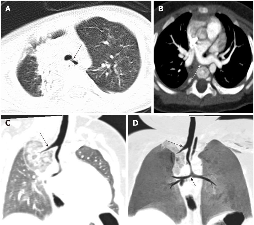
Figure 7 Tracheal bronchus with pulmonary artery sling.
Axial image in the lung window (A) shows the right upper lobe bronchus (long arrow) seen arising directly from the trachea. Axial image in mediastinal (B) shows the left pulmonary artery sling arising from the right branch pulmonary artery (long arrow). There is a separate origin of right upper lobe bronchus (long arrows) from the trachea, and the carinal angle is obtuse as seen in multiplanar reformatted images (C) and minimal intensity projection images (minip) (D) (short arrow). These findings were best depicted by minIP.
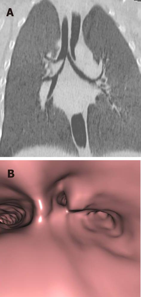
Figure 8 Tracheal trifurcation.
Coronal minimal intensity projection images (A) and virtual bronchoscopy (B) images depict tracheal trifurcation and the origin of the three bronchi, respectively.
Figure 9 Tracheal diverticulum with accessory right upper lobe bronchus.
Coronal multiplanar reformatted images in the lung window shows the presence of a blind-ending outgrowth from the left wall of the trachea (long arrow) and the presence of an accessory right upper lobe bronchus (short arrow).
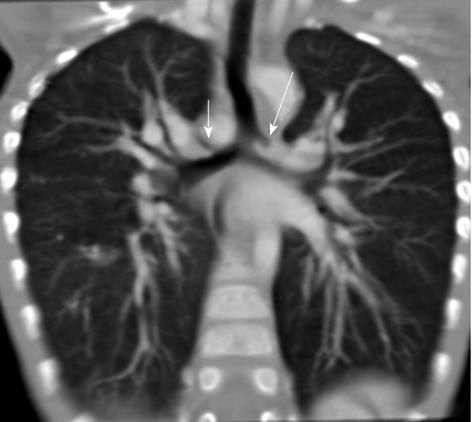
Figure 10 Isomerism.
Minimal intensity projection image (A), multiplanar reformatted images images in mediastinal window (B) show the presence of a rudimentary left upper lobe bronchus (short arrows), bronchus intermedius (long arrows) and lower lobe bronchus.
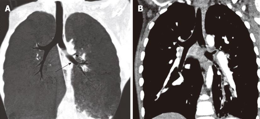
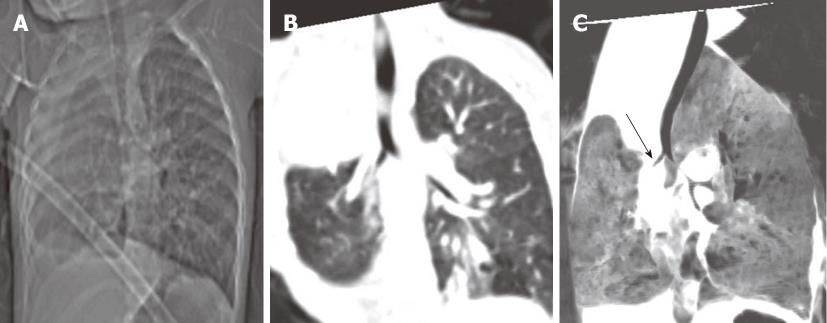
Figure 11 Congenital absence of right upper lobe and right middle lobe.
Chest radiograph (A) reveals a small right lung and coronal multiplanar reformatted images (B) and coronal minimal intensity projection images (C) show the lower lobe bronchus (arrow). The right upper lobe and right middle lobe bronchi are absent.
Figure 12 Pulmonary hypoplasia with broncho-esophageal fistula.
Axial image in the lung window (A) shows collapse of the right lower lobe. Aspirated barium (arrow) in the lower lobe was visualized in the axial image in the mediastinal window (B); C: Barium examination, carried out prior to computed tomography through an esophageal tube, depicts the origin of the right lower lobe bronchus from the esophagus.
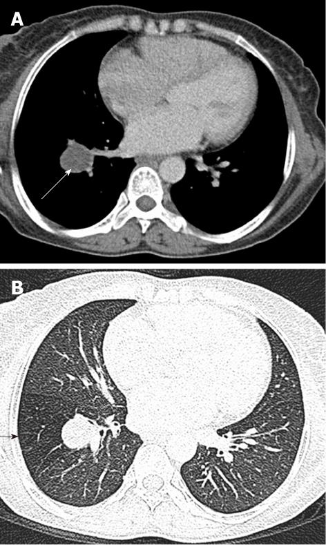
Figure 13 Bronchial atresia.
Axial image in the mediastinal window (A) shows a fluid attenuating lesion with a thin wall in the left upper lobe (long arrow). High resolution computed tomography lung window (B) image at the same level shows the presence of hyperinflation of the lung segment peripheral to the lesion (short arrow).
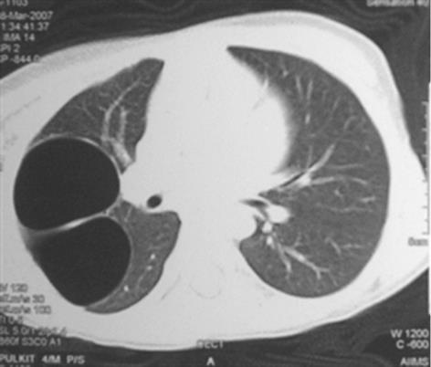
Figure 14 Congenital cystic adenomatoid malformation.
Axial image in the lung window depicts the presence of thin walled and septated air-filled cysts (arrow) in the right upper lobe, causing mass effect on the adjacent lung suggestive of congenital cystic adenomatoid malformation type-1.
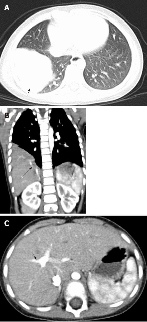
Figure 15 Sequestration of lung segment- Extralobar type.
A: Axial image in the lung window shows a homogenous pulmonary lesion in the location of the right lower lobe lateral basal segment; B, C: Coronal multiplanar reformatted images (B) and axial image (C) at the level of portal vein bifurcation in the mediastinal window show anomalous venous drainage (long arrow) into the right branch of the portal vein (short arrow).
Figure 16 Compression of left main bronchus between enlarged pulmonary artery and descending aorta.
Axial images in the mediastinal window (A and B) show enlarged pulmonary artery in this case of atrial septal defect. Axial image (B) and coronal multiplanar reformatted images in the lung window (C) show narrowing of the mid segment of the left main bronchus (arrows) and the resultant hyperinflation in the left lung.
Figure 17 Peribronchial hamartoma.
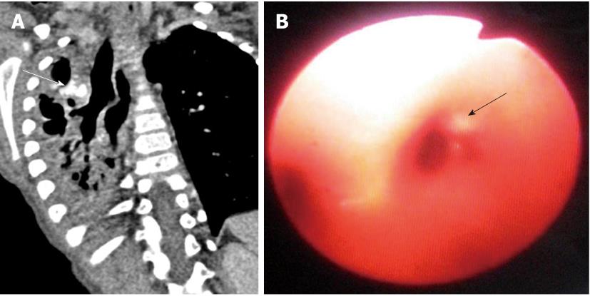
A: Coronal multiplanar reformatted images in the mediastinal window shows eccentric narrowing of the right main bronchus by a lesion with dense calcification (arrow) and resultant distal bronchiectasis and volume loss; B: Fiberoptic image of the carina shows distortion of the antero-lateral wall of the right main bronchus (arrow). Histological analysis revealed the lesion to be a peribronchial hamartoma.
- Citation: Sundarakumar DK, Bhalla AS, Sharma R, Gupta AK, Kabra SK, Jagia P. Multidetector computed tomography imaging of congenital anomalies of major airways: A pictorial essay. World J Radiol 2011; 3(12): 289-297
- URL: https://www.wjgnet.com/1949-8470/full/v3/i12/289.htm
- DOI: https://dx.doi.org/10.4329/wjr.v3.i12.289


