Copyright
©2011 Baishideng Publishing Group Co.
World J Radiol. Nov 28, 2011; 3(11): 256-265
Published online Nov 28, 2011. doi: 10.4329/wjr.v3.i11.256
Published online Nov 28, 2011. doi: 10.4329/wjr.v3.i11.256
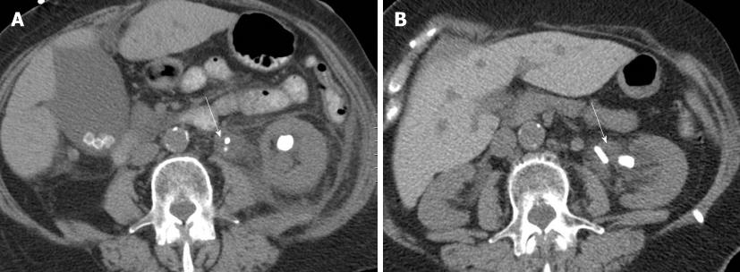
Figure 1 Transverse non-contrast abdominal computed tomography image (A) of a 78-year-old woman weighing 59 kg with left flank pain shows left renal and ureteropelvic junction stones (arrow) (14.
5 mGy, 120 kVp, 150-450 mA, noise index of 25 ) as well as multiple gallstones. On follow-up computed tomography (B), there are persistent left renal and ureteropelvic junction stones (arrow) (6.5 mGy, 120 kVp, 150-450 mA, noise index of 30).
Figure 2 Transverse non-contrast abdominal computed tomography image of a 5-year-old boy weighing 19 kg (7.
8 mGy, 120 kVp, 150-450 mA, noise index of 25) shows both renal stones (arrows) (A). Follow-up non-contrast computed tomography image obtained with 2.1 mGy, 120 kVp, 150-450 mA, and noise index of 30 shows small residual right renal stone (arrow) (B).
Figure 3 An 80-year-old man presenting with progressive increasing left flank pain and hematuria underwent abdominal computed tomography with 140 kVp and 110 mA.
A: Non-contrast transverse computed tomography (CT) image shows dilated left ureter (arrow); B: On post-contrast transverse CT image, the intra-luminal ureteral filling defect shows contrast enhancement (arrow), which was subsequently proved to be transitional cell carcinoma.
Figure 4 A 40-year-old man weighing 100 kg underwent pre-operative renal donor protocol computed tomography.
Computed tomography (CT) of the abdomen was performed with the following scan parameters; 120 kVp, 86-201 mA, 1.375 pitch and noise index of 25. Non-contrast transverse CT image (A) shows non-obstructing left renal calculus. Coronal multiplanar reformats of post-contrast CT show two right renal arteries (B) and one left renal artery (C).
- Citation: Sung MK, Singh S, Kalra MK. Current status of low dose multi-detector CT in the urinary tract. World J Radiol 2011; 3(11): 256-265
- URL: https://www.wjgnet.com/1949-8470/full/v3/i11/256.htm
- DOI: https://dx.doi.org/10.4329/wjr.v3.i11.256


