Copyright
©2010 Baishideng Publishing Group Co.
World J Radiol. Sep 28, 2010; 2(9): 345-353
Published online Sep 28, 2010. doi: 10.4329/wjr.v2.i9.345
Published online Sep 28, 2010. doi: 10.4329/wjr.v2.i9.345
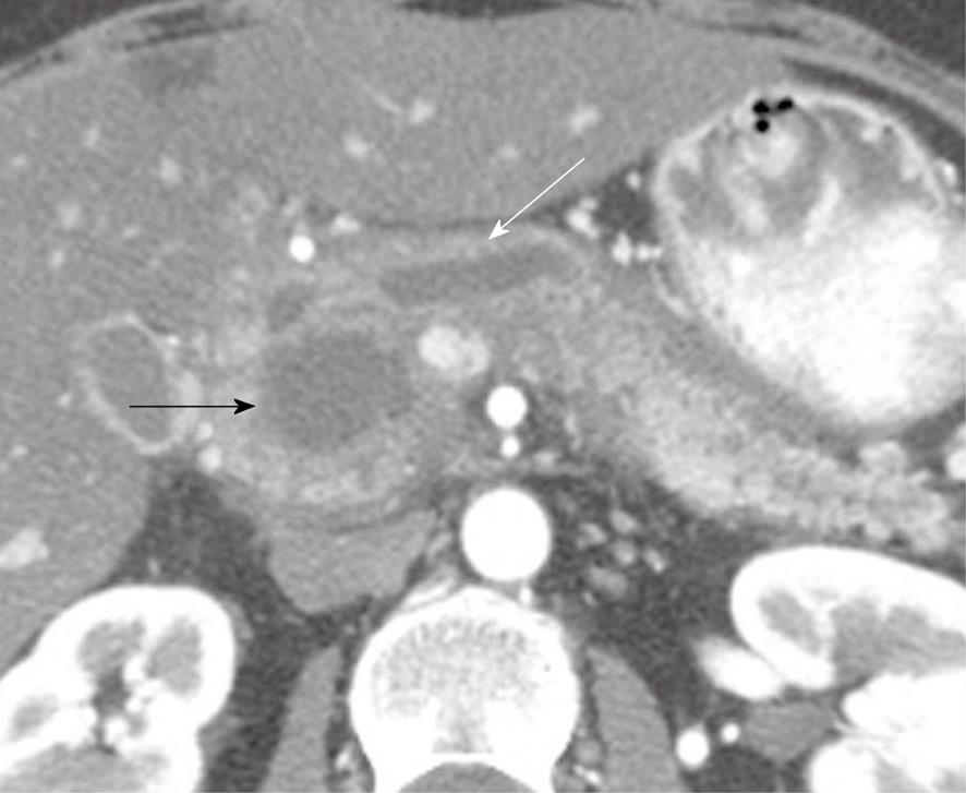
Figure 1 A 49-year-old woman with a history of epigastric and lower abdominal pain accompanied by abnormal liver function studies.
Axial contrast-enhanced computed tomography scan of the abdomen shows cystic change (black arrow) within the pancreas with associated biliary ductal dilation (white arrow). When correlated with the patient’s history and endoscopic ultrasound with FNA findings, this was consistent with a pseudocyst.
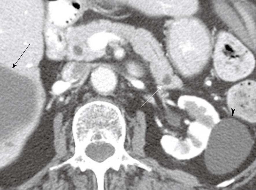
Figure 2 An 80-year-old woman with a history of hypertension, congestive heart failure and colon cancer.
Axial contrast-enhanced computed tomography of the abdomen shows a small low attenuation lesion in the pancreatic tail consistent with a cyst (white arrow), a cyst within the kidney (arrowhead) and a cyst within the liver (black arrow).
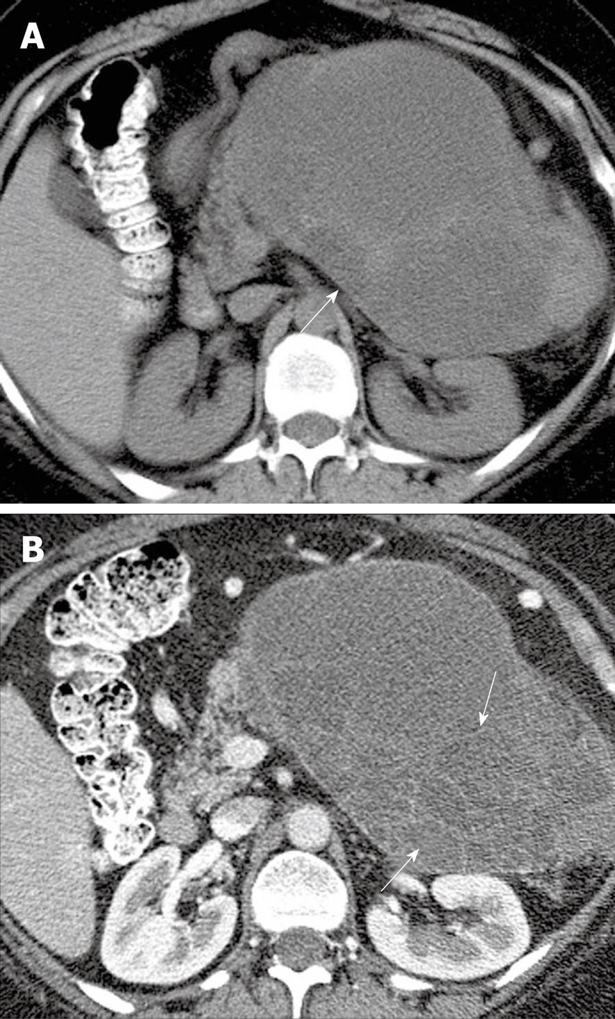
Figure 3 A 48-year-old woman with left upper quadrant pain.
A: Axial non-contrast computed tomography (CT) scan of the abdomen shows a heterogeneous mass (arrow) in the left upper quadrant; B: Axial contrast-enhanced CT of the abdomen shows enhancing septa (arrows) within the mass.
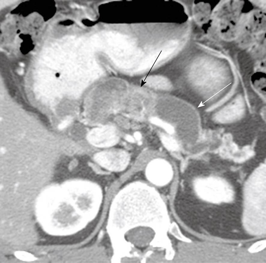
Figure 4 A 48-year-old man presenting with pancreatitis and exocrine pancreatic insufficiency and a mass in the pancreas representing diffuse intraductal papillary mucinous neoplasm of the pancreas.
Axial contrast-enhanced computed tomography of the abdomen shows dilated main pancreatic duct (white arrow) due to mucin production and an enhancing mass in the main pancreatic duct (black arrow) representing a main duct intraductal papillary mucinous neoplasm.
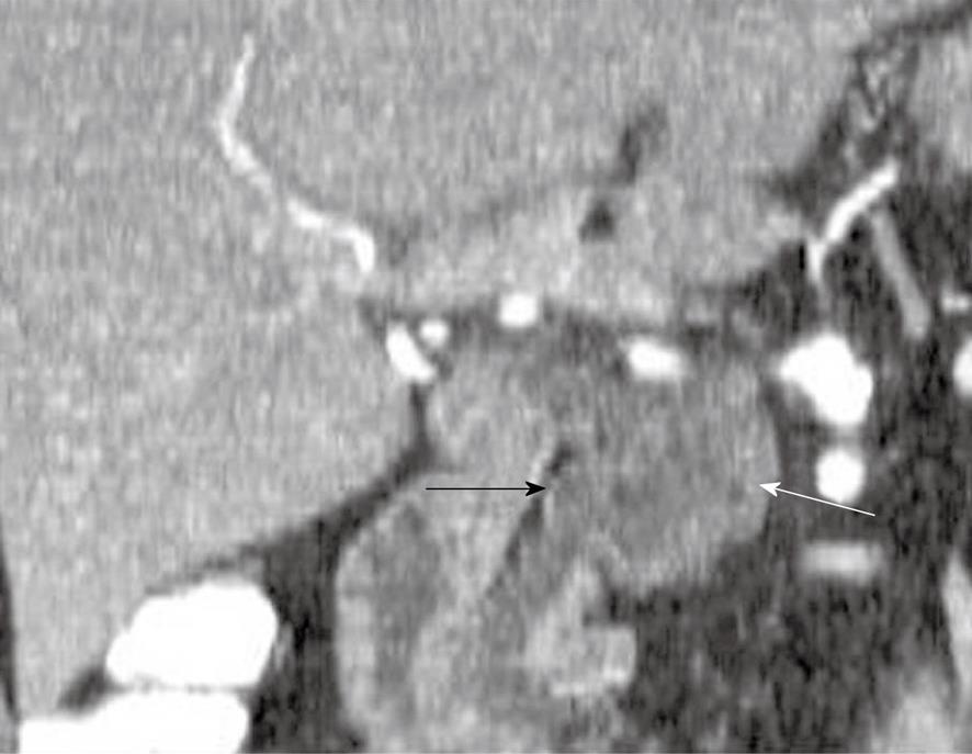
Figure 5 A 57-year-old man with cysts in the pancreas.
Coronal reformatted contrast-enhanced computed tomography scan of the abdomen shows a cystic lesion in the pancreas (white arrow) which communicates with the main pancreatic duct (black arrow).
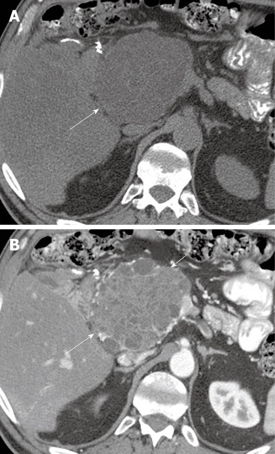
Figure 6 A 62-year-old man with history of epigastric discomfort.
A: Axial non-contrast computed tomography (CT) of the abdomen shows a mass in the pancreatic head (long arrow); B: Axial contrast-enhanced CT of the abdomen shows enhancement within the mass (long arrow) and cystic components (short arrow) suggestive of a microcystic serous cystadenoma.
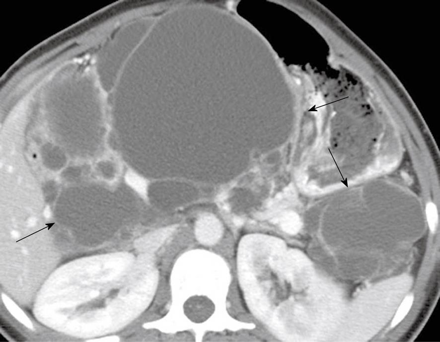
Figure 7 A 26-year-old woman with a history of von Hippel-Lindau syndrome.
Axial contrast-enhanced computed tomography scan shows cystic lesions in the pancreas (black arrows) representing oligocystic serous cyst adenomas.
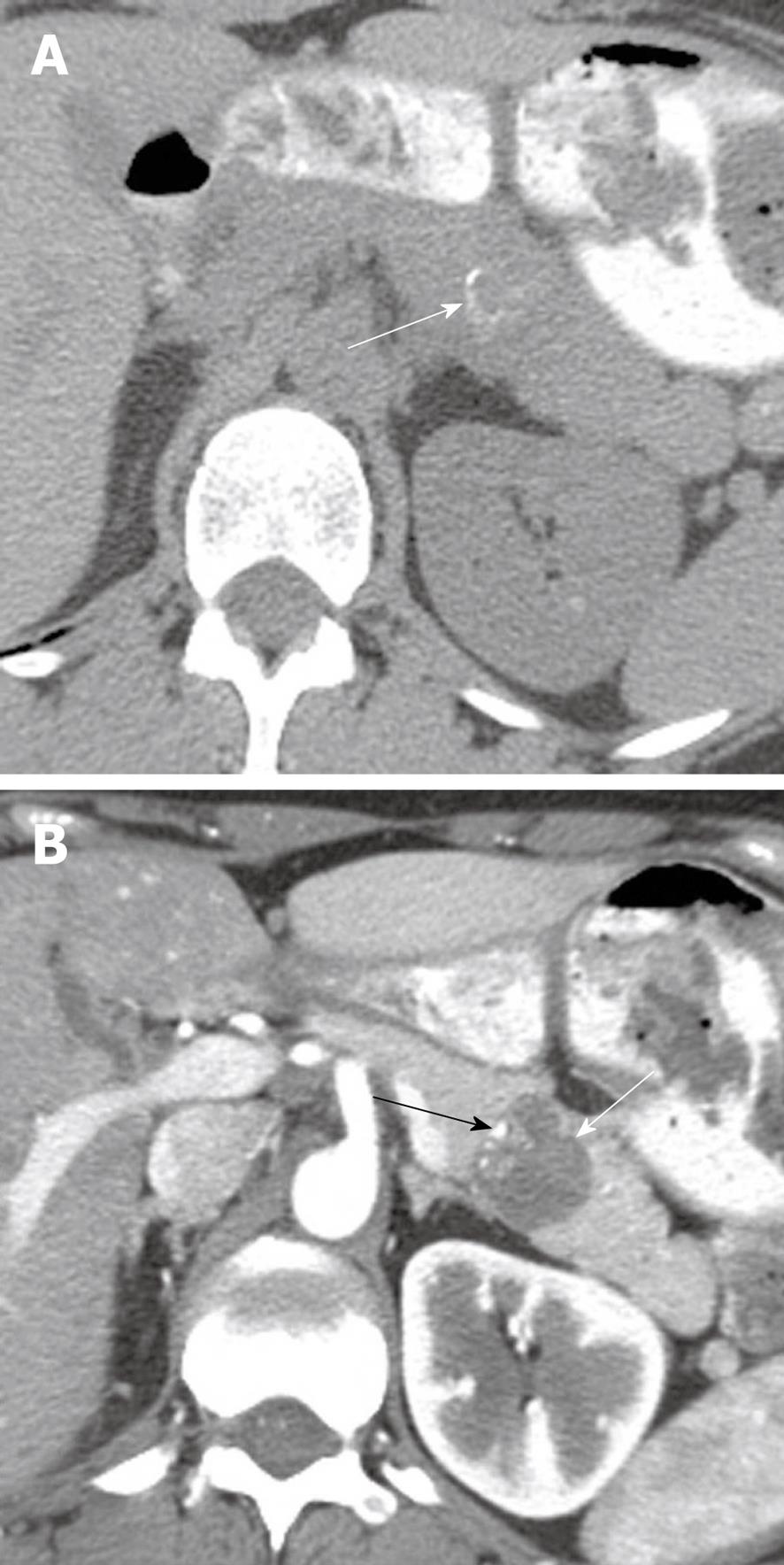
Figure 8 A 30-year-old woman with increasing abdominal discomfort and bloating.
A: Axial non contrast computed tomography (CT) of the abdomen shows a solid mass in the pancreatic tail containing curvilinear calcification (arrow); B: Axial contrast-enhanced CT of the abdomen shows a hypoattenuating mass (white arrow) in the pancreas containing eccentric calcifications (black arrow) consistent with a solid papillary epithelial neoplasm.
- Citation: Bhosale P, Balachandran A, Tamm E. Imaging of benign and malignant cystic pancreatic lesions and a strategy for follow up. World J Radiol 2010; 2(9): 345-353
- URL: https://www.wjgnet.com/1949-8470/full/v2/i9/345.htm
- DOI: https://dx.doi.org/10.4329/wjr.v2.i9.345


