Copyright
©2010 Baishideng Publishing Group Co.
World J Radiol. Sep 28, 2010; 2(9): 339-344
Published online Sep 28, 2010. doi: 10.4329/wjr.v2.i9.339
Published online Sep 28, 2010. doi: 10.4329/wjr.v2.i9.339
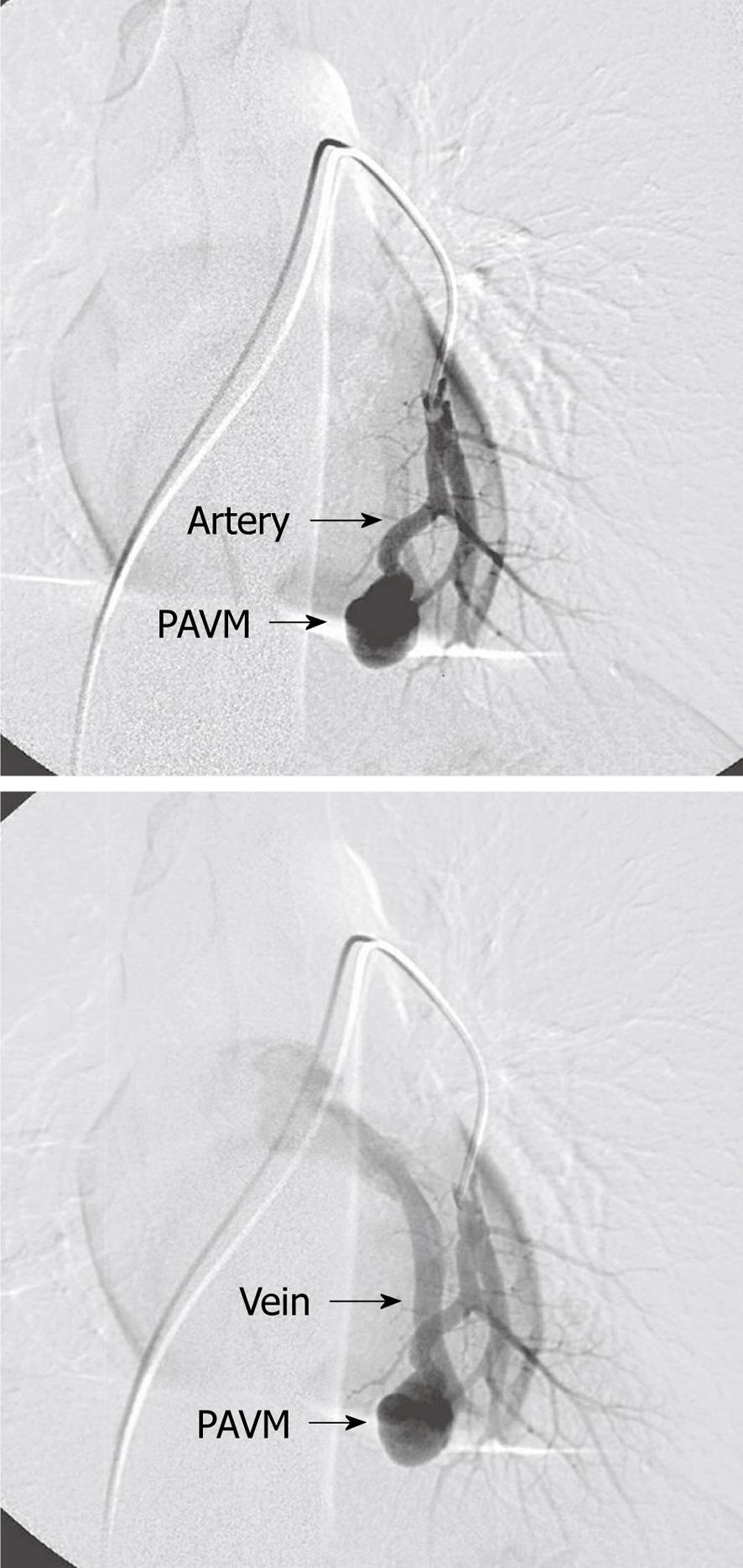
Figure 1 Simple pulmonary arteriovenous malformations with one afferent (feeding) artery and one efferent (draining) vein.
PAVM: Pulmonary arteriovenous malformations.
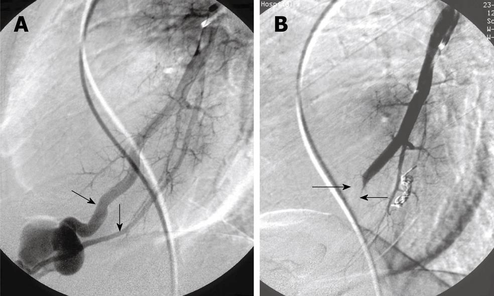
Figure 2 Complex pulmonary arteriovenous malformations.
A: Two afferent (feeding) arteries (arrows); B: After embolization with balloon in one feeding artery (arrows) and coils in the other.
Figure 3 Multiple pulmonary arteriovenous malformations.
A: In both lungs; B: Multiple pulmonary arteriovenous malformations (PAVM) after embolization. One small PAVM on the right side has been left untreated because of small sized feeding artery (arrow).
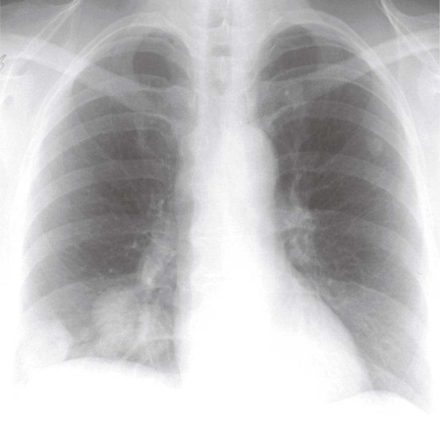
Figure 4 Pulmonary arteriovenous malformations in both lungs, visible on the chest X-ray.
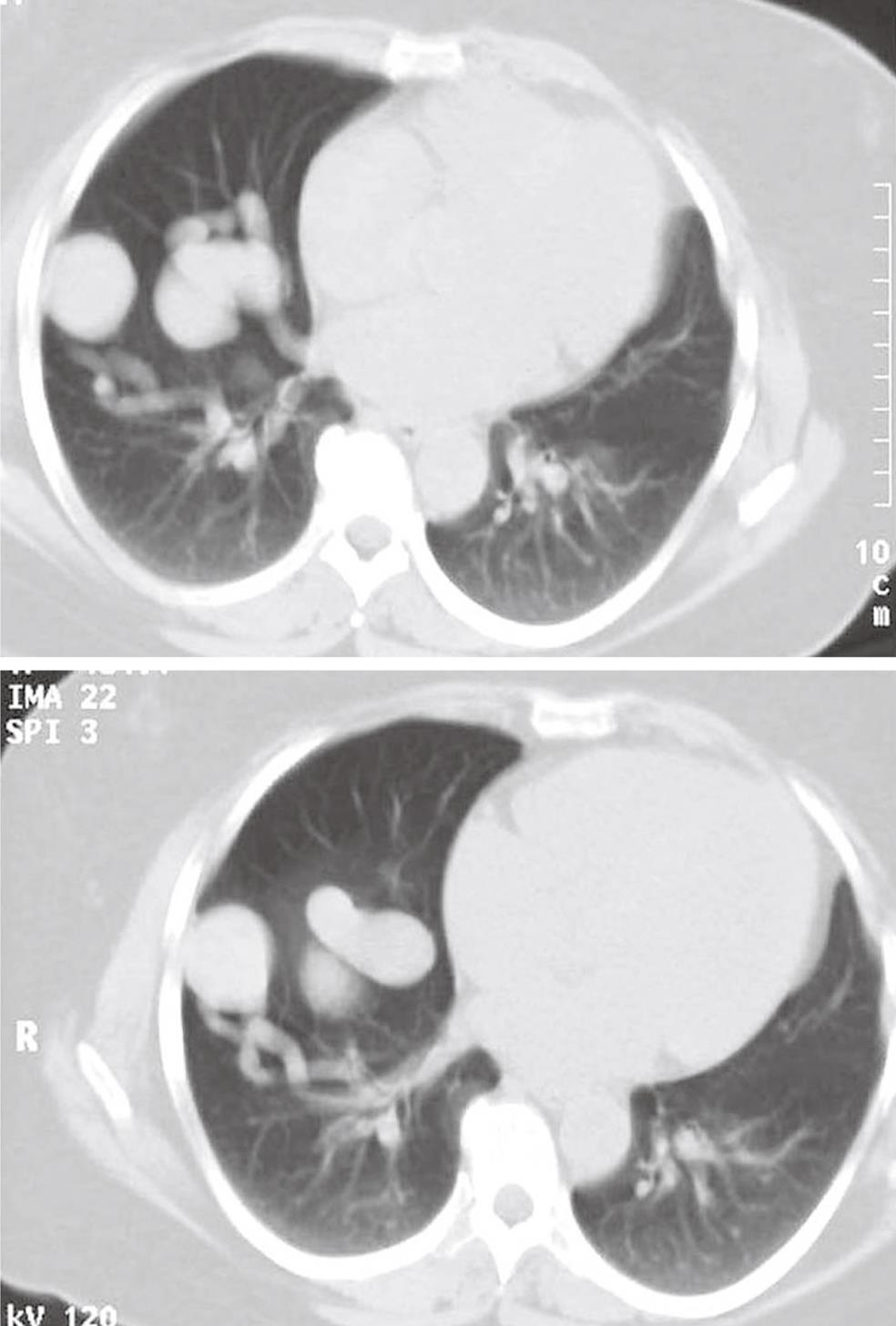
Figure 5 Computed tomography of the chest showing pulmonary arteriovenous malformations with afferent and efferent vessels.
Figure 6 Simple pulmonary arteriovenous malformations.
A, B: Before embolization; C: After selective catheterization of the feeding artery; D: After embolization with coils (arrow).
Figure 7 Embolization of simple pulmonary arteriovenous malformations with vascular plug.
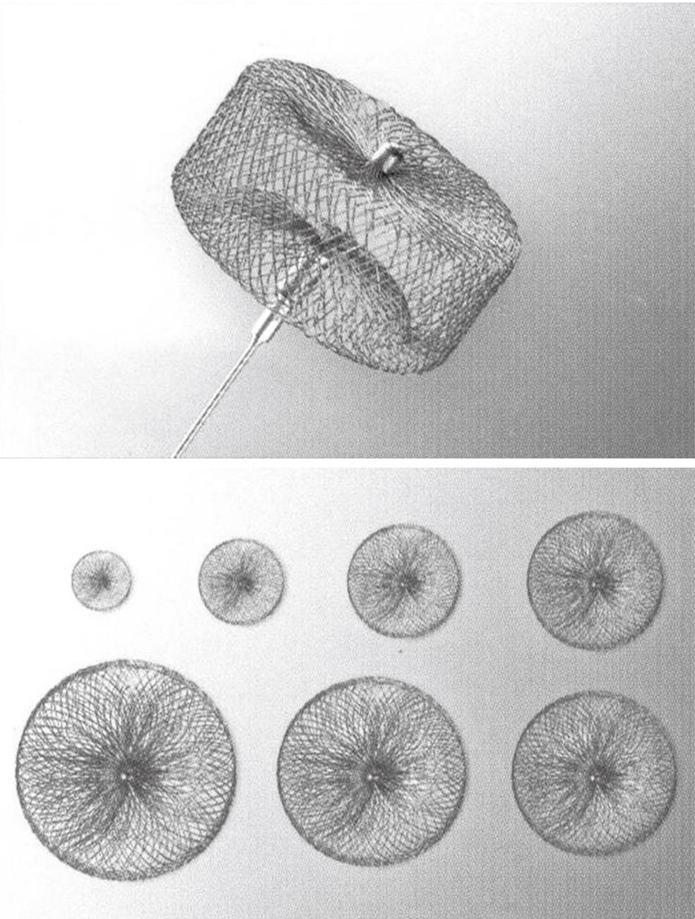
Figure 8 One version of a vascular plug.
- Citation: Andersen PE, Kjeldsen AD. Interventional treatment of pulmonary arteriovenous malformations. World J Radiol 2010; 2(9): 339-344
- URL: https://www.wjgnet.com/1949-8470/full/v2/i9/339.htm
- DOI: https://dx.doi.org/10.4329/wjr.v2.i9.339


