Copyright
©2010 Baishideng Publishing Group Co.
World J Radiol. Jul 28, 2010; 2(7): 257-261
Published online Jul 28, 2010. doi: 10.4329/wjr.v2.i7.257
Published online Jul 28, 2010. doi: 10.4329/wjr.v2.i7.257
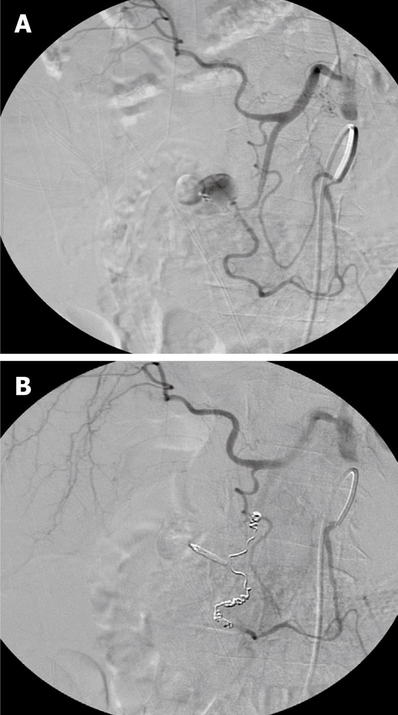
Figure 1 Ninety years old female with severe acute duodenal ulcerous bleeding.
A: Primary endoscopic treatment but suffered rebleeding and was remitted for endovascular treatment. Bleeding from gastroduodenal artery; B: Bleeding stopped after embolization with coils of gastroduodenal artery distally and proximally to the former bleeding part of the artery.
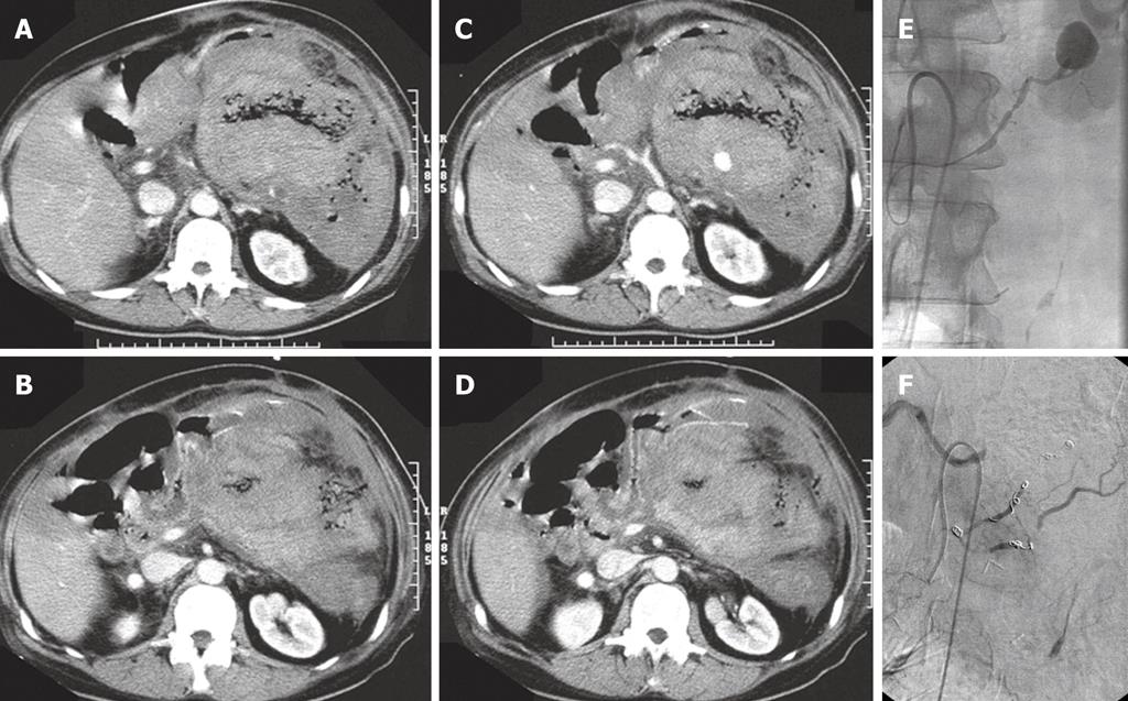
Figure 2 Fifty-four years old male with hereditary polyposis and colectomy some years earlier.
A-D: Because of polyps in the second part of the duodenum, Whipple’s operation was performed. Two weeks later severe upper gastrointestinal bleeding was seen. Abdominal computed tomography shows a large intraperitoneal haematoma and active bleeding and/or (pseudo)aneurysm; E: Superior mesenteric arteriography showing pseudoaneurysm and bleeding from, most probably, a transverse pancreatic artery branch. Vascular spasms are seen after placement of a microcatheter in the vessel; F: After embolization with microcoils in two branches supplying the pseudoaneurysm/bleeding.
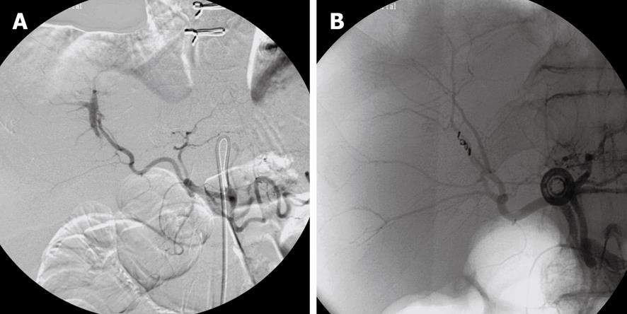
Figure 3 Sixty-four years old male with aortobiliacal prosthesis 2 years earlier.
A: Computed tomography performed because of abdominal pain. The liver appeared with parenchymatous changes and large amounts of ascites. Percutaneous liver biopsy was performed following hematemesis and melaena and bleeding from the papilla seen at endoscopy. Selective celiac angiography shows fistula from right hepatic artery branch to bile duct; B: Arteriobiliary fistula occluded after embolization with micro coils.
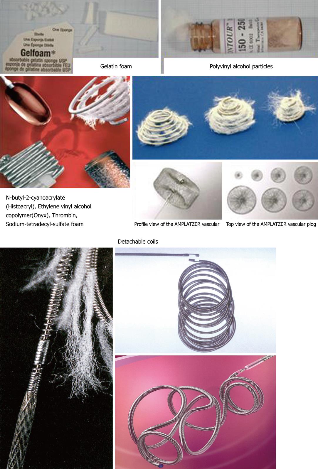
Figure 4 Examples of different embolization materials.
- Citation: Andersen PE, Duvnjak S. Endovascular treatment of nonvariceal acute arterial upper gastrointestinal bleeding. World J Radiol 2010; 2(7): 257-261
- URL: https://www.wjgnet.com/1949-8470/full/v2/i7/257.htm
- DOI: https://dx.doi.org/10.4329/wjr.v2.i7.257


