Copyright
©2010 Baishideng Publishing Group Co.
World J Radiol. May 28, 2010; 2(5): 193-196
Published online May 28, 2010. doi: 10.4329/wjr.v2.i5.193
Published online May 28, 2010. doi: 10.4329/wjr.v2.i5.193
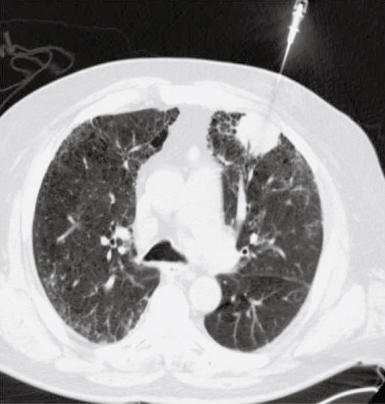
Figure 1 Chest computed tomography (CT) demonstrates the left upper lobe mass during the biopsy.
The mass abuts the chest wall with mild adjacent emphysematous changes without large bullae.
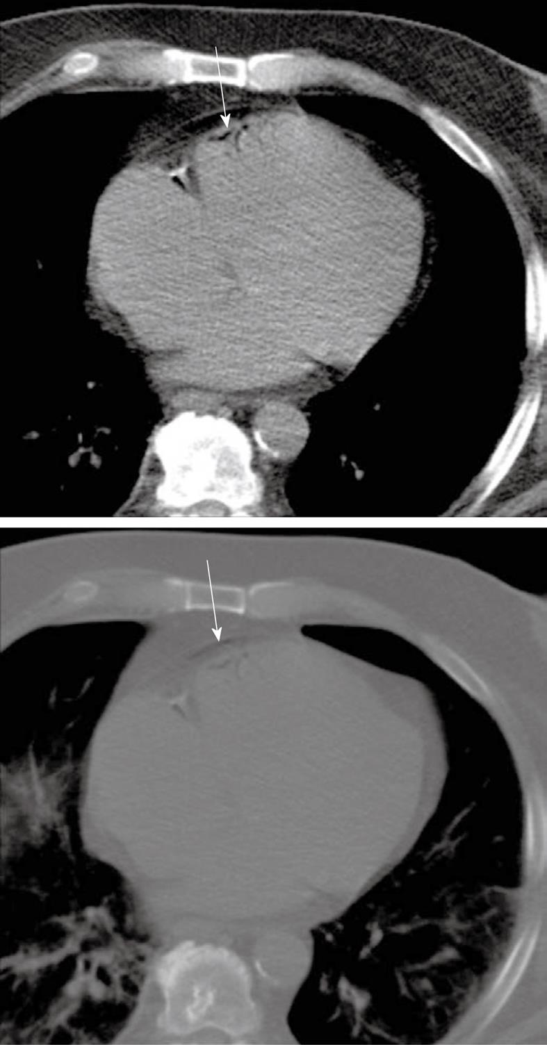
Figure 2 Axial images in soft tissue and lung window through the lower mediastinum, display a small amount of air in the epicardium (arrows), thought to be along the peripheral coronary vessels.
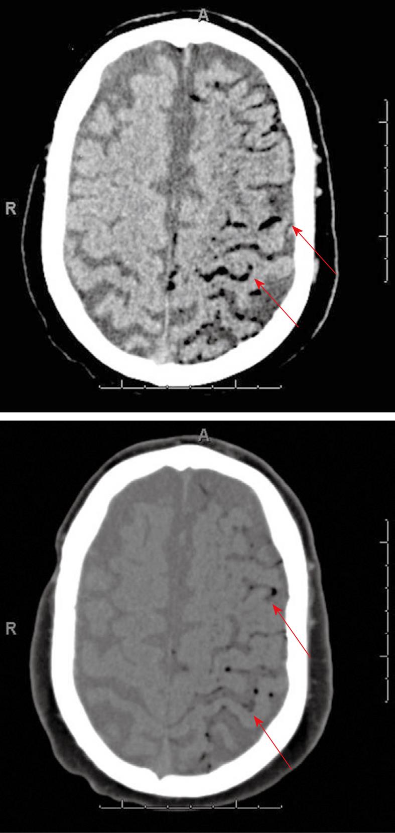
Figure 3 Head CT in brain and soft tissue windows demonstrates abnormal air along the left vertex subarachnoid spaces and cortical vessels (arrows), suggesting air embolus.
- Citation: Bou-Assaly W, Pernicano P, Hoeffner E. Systemic air embolism after transthoracic lung biopsy: A case report and review of literature. World J Radiol 2010; 2(5): 193-196
- URL: https://www.wjgnet.com/1949-8470/full/v2/i5/193.htm
- DOI: https://dx.doi.org/10.4329/wjr.v2.i5.193


