Copyright
©2010 Baishideng Publishing Group Co.
World J Radiol. May 28, 2010; 2(5): 188-192
Published online May 28, 2010. doi: 10.4329/wjr.v2.i5.188
Published online May 28, 2010. doi: 10.4329/wjr.v2.i5.188
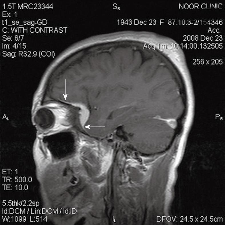
Figure 1 A 65-year-old woman with meningioma and adjacent hyperostosis.
Arrows indicates “dural tail sign”.
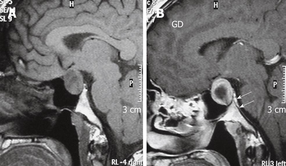
Figure 2 Imaging findings of pituitary macroadenoma.
A: T1W sagittal MRI of brain shows a Pituitary macro-adenoma with extension to the suprasellar cistern; B: After gadolinium injection enhancement is noted in periphery of tumour and dura of dorsum sella (arrows), enhancement is greater than that of the tumour itself. Biopsy proved prolactinoma.
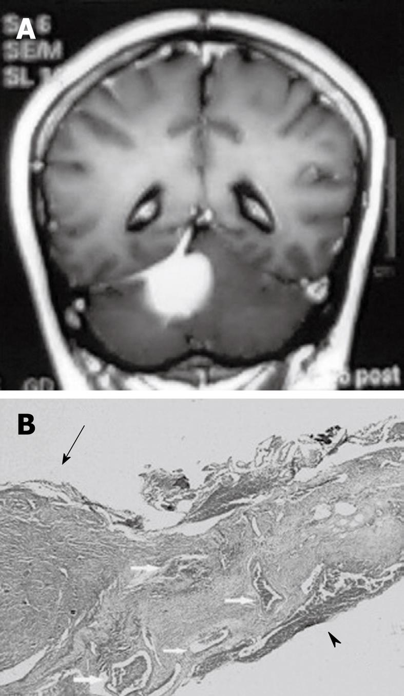
Figure 3 Pathological finding of dural tail sign.
A: A 58-year-old female with a tentorial meningioma and dural tail sign; B: Pathology specimen in this case showed a meningioma (arrow) and the attached dura. The dural tail is noted on the right side of the figure (arrowhead). Both the dura beneath the tumor and dural tail contain dilated blood vessels (arrow). There is no dural invasion. HE 30 ×.
- Citation: Sotoudeh H, Yazdi HR. A review on dural tail sign. World J Radiol 2010; 2(5): 188-192
- URL: https://www.wjgnet.com/1949-8470/full/v2/i5/188.htm
- DOI: https://dx.doi.org/10.4329/wjr.v2.i5.188


